Degradation of deproteinized natural rubber by Gordonia sp. isolated from enrichment consortia
Biodegradation is a potential way of decomposing deproteinized natural rubber
(DPNR). The enrichment consortia were demonstrated from a rubber processing factory waste.
Nine DPNR-degrading bacteria were isolated from those consortia. The highest DPNR film
weight loss in a mineral salt medium (MSM) was 43.92 ± 2.30 % after 30 days incubation using
strain 5A1. The formation of aldehyde group during rubber degradation of 5A1 was determined
using Schiff staining and Fourier Transform Infrared spectroscopy (FTIR) analysis. The 16S
rRNA gene sequence, of 5A1 showed the highest identity with that of Gordonia soli CC-AB07.
This is the first report to demonstrate a strong ability to degrade DPNR by Gordonia sp. isolated
from a rubber processing factory waste in Viet Nam
Trang 1
Trang 2
Trang 3
Trang 4
Trang 5
Trang 6
Trang 7
Trang 8
Tóm tắt nội dung tài liệu: Degradation of deproteinized natural rubber by Gordonia sp. isolated from enrichment consortia
-overlay agar
plates for further study.
 The individual isolated strain was incubated in 20 mL of MSM supplemented with DPNR
films (0.3 %, w/v) as a sole carbon source at 30 ◦C, 150 rpm for 30 days. Each experiment was
performed in duplicate. After incubation, DPNR films were collected and washed with 0.1 M
NaOH for 30 minutes followed by distilled water to remove attached biomass. Samples were
then dried at 50 °C until a constant weight [3]. The weight loss of DPNR films was measured
before (W1) and after degradation (W2). Weight loss (%) was calculated as follows:
 Weight loss = x 100
2.2.2. Deproteinized natural rubber degradation by selected isolate
 The selected strain was obtained based on the highest DPNR film weight loss after 30 days
incubation. It was grown on a DPNR-overlay plate after 7 days and stained with Schiff’s
reagent. The purple colour produced by the reagent shows the evidence that polyisoprene
oligomers containg aldehyde group was accumulated during the microbial degradation of rubber.
Staining was performed as described previously [9].
 The FTIR (Jasco-4600, Japan) analysis was done to detect the degradation of DPNR film
after 30 days inculation by selected strain in MSM at 30 oC and 150 rpm on the basis of changes
in the functional group. The DPNR films was mixed with KBr and made into a pellet, which was
fixed to the FTIR sample plate. Spectra were taken at 400 to 4000 cm–1 wave numbers for each
sample. The correlation of absorption bands in the spectrum of an unknown compound with the
known absorption frequencies were analyzed [10].
2.2.3. 16S RNA gene analysis
 The total genomic DNA of the selected strain was extracted using a QIAamp DNA mini kit
(QIAGEN). The 16S rRNA gene sequences were amplified using the bacterial universal primers
named 27F (5’-AGAGTTTGATCCTGGCTCAG-3’) and 1492R (5'-
GGTTACCTTGTTACGACTT-3') [21]. The PCR reaction was carried out in a final volume of
20 µl containing 10 µl PCR Master mix 2x Promega Taq DNA Polymerase, 1 µl primer 10 µM
27F, 1 µl primer 10 µM 1492R, 1 µl of the DNA template and 7 µl MiliQ. The PCR was
performed according to the following program: 2 min denaturation at 94 oC, followed by 30
cycles of 1 min denaturation at 94 oC; 1 min annealing at 50 oC; 1 min extension at 72 oC; and a
final extension step of 2 min at 72 oC. The 16S rDNA gene was sequenced. The sequences
analysis was carried out on an ABI Prism, 3100-Avant Genetic Analyzer (Hitachi, Japan).
 The gene sequences were aligned with published sequence in Genbank database using the
basic local alignment search tool (BLAST) at the United State National Center of Biotechnology
86
Finite element modelling for electric field distribution around positive streamers in oil
Information. A phylogenetic analysis was performed with the Clustal W program using the
neighbour joining method.
2.2.4. Nucleotide sequence accession number
 The partial 16S rRNA nucleotide sequence of the selected strain has been deposited in the
GenBank database.
 3. RESULTS AND DISCUSSION
3.1. Screening of deproteinized natural rubber degrading bacteria
 To enhance the growth of deproteinized natural rubber degrading bacteria, the enrichment
consortia were demonstrated with five sub-transfers. The nine isolates obtained from enrichment
consortia were able to grow on MSM agar containing DPNR as the sole carbon source. Only one
of isolate, 1A2 formed a clearing zone around the colony and other isolates did not show the
clearing zone around the colonies. However, all isolates grew well in the liquid MSM with
DPNR films. The degradation of rubber by isolates was determined based on the DPNR film
weight loss in Figure 1.
 Figure 1. DPNR film weight loss after 30 days incubation with isolates.
 After 30 days incubation, the weight loss of DPNR film by isolates varied from 9.31 ± 1.32
to 43.92 ± 2.30 %. The highest weight loss was obtained from the culture of strain 5A1.
Nawong C. et al. isolated Rhodococcus pyridinivorans strain F5 from soil samples in Thailand,
the weight loss of latex glove pieces for 30 days of incubation by F5 at 30oC was 9.36 % [10]. In
another study Gallert C. showed the weight loss of latex gloves by Streptomyces sp. strain La7
for 32 days of incubation at 30 oC was 13.5 % [11]. Trang N. et al. reported that four NR
degrading strains were isolated from waste in Cam Thuy of Viet Nam belonging to Streptomyces
sp. The weight loss of NR pieces of strain NMD1, strain M4D1b, strain M4T1c, and strain T37
at 30 oC for 30 days were 4.28 ± 0.32 %, 3.11 ± 0.5 %, 2.05 ± 0.76 % and 2.43 ± 0.34 %,
respectively [12]. It seems that strain 5A1 had strong ability to degrade DPNR.
3.2. Deproteinized natural rubber degrading bacteria by strain 5A1
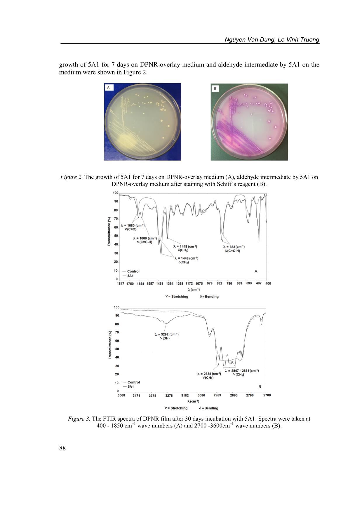
 In order to determine the rubber degradation products contain aldehyde groups, the
acumulation of aldehydes on DPNR-overlay agar plate was examined by Schiff’s reagent. The
 87
 Nguyen Van Dung, Le Vinh Truong
growth of 5A1 for 7 days on DPNR-overlay medium and aldehyde intermediate by 5A1 on the
medium were shown in Figure 2.
Figure 2. The growth of 5A1 for 7 days on DPNR-overlay medium (A), aldehyde intermediate by 5A1 on
 DPNR-overlay medium after staining with Schiff’s reagent (B).
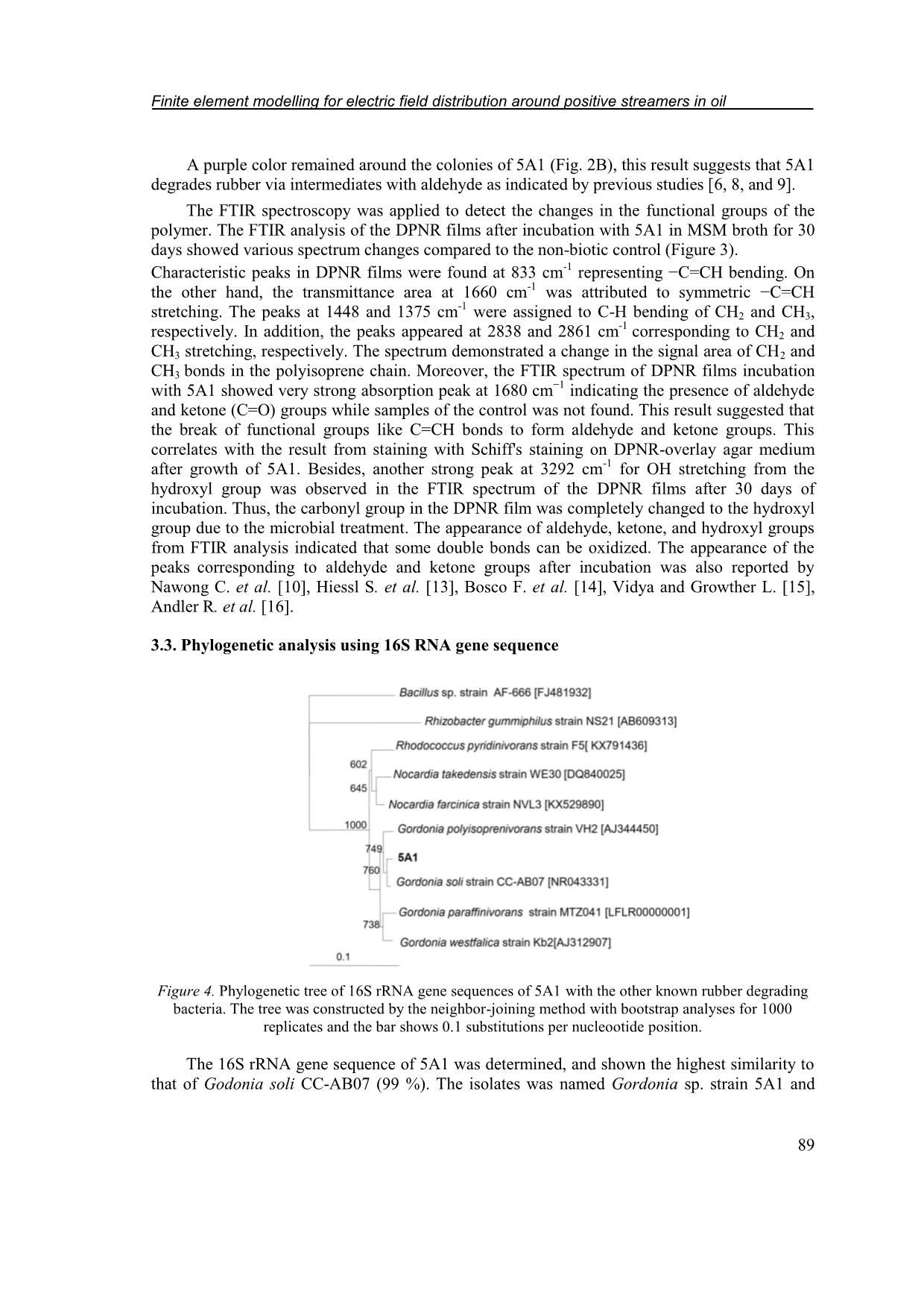
 Figure 3. The FTIR spectra of DPNR film after 30 days incubation with 5A1. Spectra were taken at
 400 - 1850 cm–1 wave numbers (A) and 2700 -3600cm–1 wave numbers (B).
88
Finite element modelling for electric field distribution around positive streamers in oil
 A purple color remained around the colonies of 5A1 (Fig. 2B), this result suggests that 5A1
degrades rubber via intermediates with aldehyde as indicated by previous studies [6, 8, and 9].
 The FTIR spectroscopy was applied to detect the changes in the functional groups of the
polymer. The FTIR analysis of the DPNR films after incubation with 5A1 in MSM broth for 30
days showed various spectrum changes compared to the non-biotic control (Figure 3).
Characteristic peaks in DPNR films were found at 833 cm-1 representing −C=CH bending. On
the other hand, the transmittance area at 1660 cm-1 was attributed to symmetric −C=CH
 -1
stretching. The peaks at 1448 and 1375 cm were assigned to C-H bending of CH2 and CH3,
 -1
respectively. In addition, the peaks appeared at 2838 and 2861 cm corresponding to CH2 and
CH3 stretching, respectively. The spectrum demonstrated a change in the signal area of CH2 and
CH3 bonds in the polyisoprene chain. Moreover, the FTIR spectrum of DPNR films incubation
with 5A1 showed very strong absorption peak at 1680 cm−1 indicating the presence of aldehyde
and ketone (C=O) groups while samples of the control was not found. This result suggested that
the break of functional groups like C=CH bonds to form aldehyde and ketone groups. This
correlates with the result from staining with Schiff's staining on DPNR-overlay agar medium
after growth of 5A1. Besides, another strong peak at 3292 cm-1 for OH stretching from the
hydroxyl group was observed in the FTIR spectrum of the DPNR films after 30 days of
incubation. Thus, the carbonyl group in the DPNR film was completely changed to the hydroxyl
group due to the microbial treatment. The appearance of aldehyde, ketone, and hydroxyl groups
from FTIR analysis indicated that some double bonds can be oxidized. The appearance of the
peaks corresponding to aldehyde and ketone groups after incubation was also reported by
Nawong C. et al. [10], Hiessl S. et al. [13], Bosco F. et al. [14], Vidya and Growther L. [15],
Andler R. et al. [16].
3.3. Phylogenetic analysis using 16S RNA gene sequence
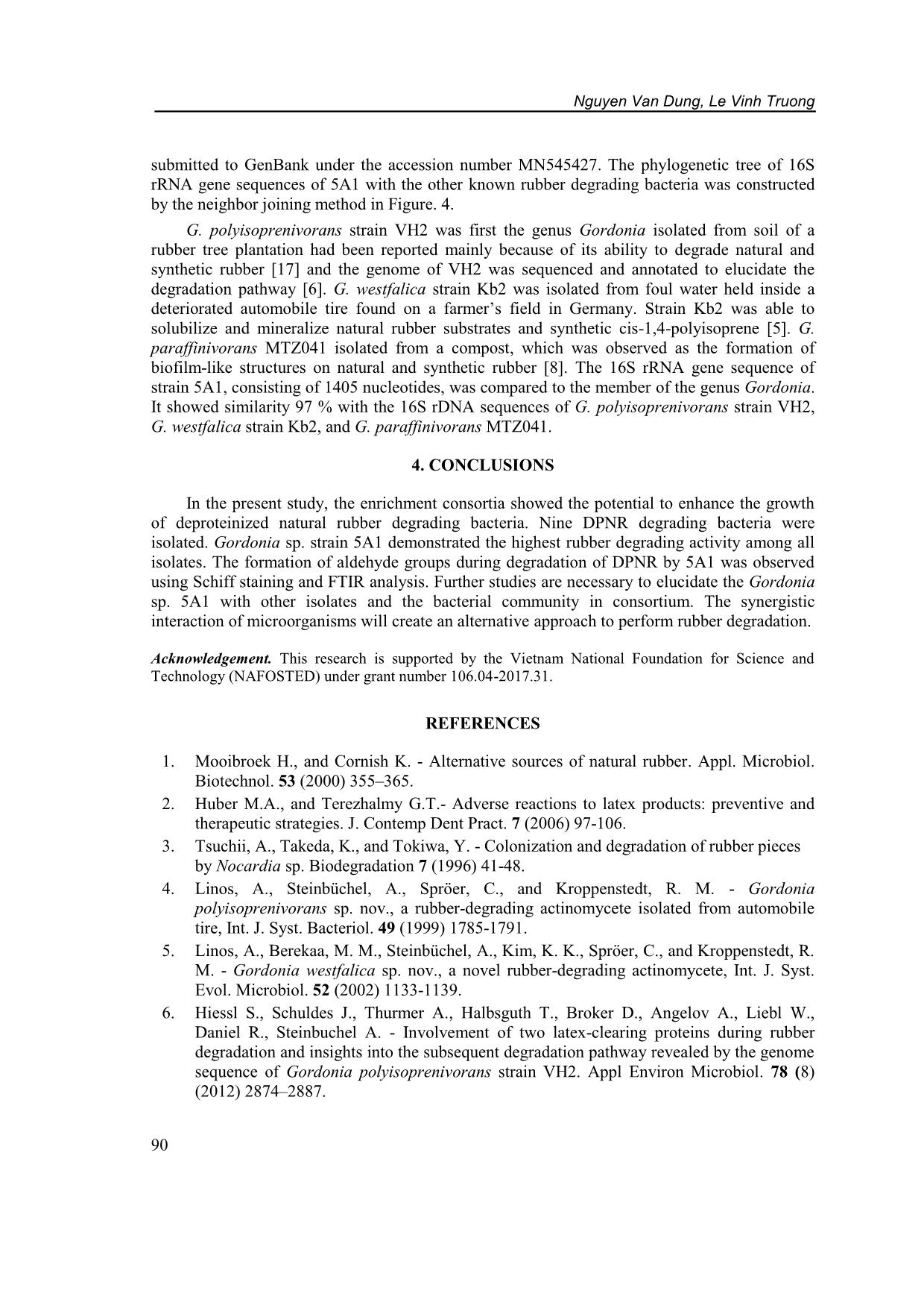
Figure 4. Phylogenetic tree of 16S rRNA gene sequences of 5A1 with the other known rubber degrading
 bacteria. The tree was constructed by the neighbor-joining method with bootstrap analyses for 1000
 replicates and the bar shows 0.1 substitutions per nucleootide position.
 The 16S rRNA gene sequence of 5A1 was determined, and shown the highest similarity to
that of Godonia soli CC-AB07 (99 %). The isolates was named Gordonia sp. strain 5A1 and
 89
 Nguyen Van Dung, Le Vinh Truong
submitted to GenBank under the accession number MN545427. The phylogenetic tree of 16S
rRNA gene sequences of 5A1 with the other known rubber degrading bacteria was constructed
by the neighbor joining method in Figure. 4.
 G. polyisoprenivorans strain VH2 was first the genus Gordonia isolated from soil of a
rubber tree plantation had been reported mainly because of its ability to degrade natural and
synthetic rubber [17] and the genome of VH2 was sequenced and annotated to elucidate the
degradation pathway [6]. G. westfalica strain Kb2 was isolated from foul water held inside a
deteriorated automobile tire found on a farmer’s field in Germany. Strain Kb2 was able to
solubilize and mineralize natural rubber substrates and synthetic cis-1,4-polyisoprene [5]. G.
paraffinivorans MTZ041 isolated from a compost, which was observed as the formation of
biofilm-like structures on natural and synthetic rubber [8]. The 16S rRNA gene sequence of
strain 5A1, consisting of 1405 nucleotides, was compared to the member of the genus Gordonia.
It showed similarity 97 % with the 16S rDNA sequences of G. polyisoprenivorans strain VH2,
G. westfalica strain Kb2, and G. paraffinivorans MTZ041.
 4. CONCLUSIONS
 In the present study, the enrichment consortia showed the potential to enhance the growth
of deproteinized natural rubber degrading bacteria. Nine DPNR degrading bacteria were
isolated. Gordonia sp. strain 5A1 demonstrated the highest rubber degrading activity among all
isolates. The formation of aldehyde groups during degradation of DPNR by 5A1 was observed
using Schiff staining and FTIR analysis. Further studies are necessary to elucidate the Gordonia
sp. 5A1 with other isolates and the bacterial community in consortium. The synergistic
interaction of microorganisms will create an alternative approach to perform rubber degradation.
Acknowledgement. This research is supported by the Vietnam National Foundation for Science and
Technology (NAFOSTED) under grant number 106.04-2017.31.
 REFERENCES
 1. Mooibroek H., and Cornish K. - Alternative sources of natural rubber. Appl. Microbiol.
 Biotechnol. 53 (2000) 355–365.
 2. Huber M.A., and Terezhalmy G.T.- Adverse reactions to latex products: preventive and
 therapeutic strategies. J. Contemp Dent Pract. 7 (2006) 97-106.
 3. Tsuchii, A., Takeda, K., and Tokiwa, Y. - Colonization and degradation of rubber pieces
 by Nocardia sp. Biodegradation 7 (1996) 41-48.
 4. Linos, A., Steinbüchel, A., Spröer, C., and Kroppenstedt, R. M. - Gordonia
 polyisoprenivorans sp. nov., a rubber-degrading actinomycete isolated from automobile
 tire, Int. J. Syst. Bacteriol. 49 (1999) 1785-1791.
 5. Linos, A., Berekaa, M. M., Steinbüchel, A., Kim, K. K., Spröer, C., and Kroppenstedt, R.
 M. - Gordonia westfalica sp. nov., a novel rubber-degrading actinomycete, Int. J. Syst.
 Evol. Microbiol. 52 (2002) 1133-1139.
 6. Hiessl S., Schuldes J., Thurmer A., Halbsguth T., Broker D., Angelov A., Liebl W.,
 Daniel R., Steinbuchel A. - Involvement of two latex-clearing proteins during rubber
 degradation and insights into the subsequent degradation pathway revealed by the genome
 sequence of Gordonia polyisoprenivorans strain VH2. Appl Environ Microbiol. 78 (8)
 (2012) 2874–2887.
90
Finite element modelling for electric field distribution around positive streamers in oil
 7. Berekaa M. M., Lino A., Reichelt R., Keller U., and Steinbuchel A. - Effect of
 pretreatment of rubbber material on its biodegradability by various rubber degrading
 bacteria. FEMS Microbiol. Lett. 184 (2000) 199-206.
 8. Braga S. P., Santos, A. P., Paganini T., Barbosa D Epamino G. W. C., Morais C., Martins
 L. F., Silva A. M., Setubal J. C., Vallim M. A., and Pascon R. C. - First report of cis-1,4-
 polyisoprene degradation by Gordonia paraffinivorans. Brazilian Journal of
 Microbiology, 2019, https://doi.org/10.1007/s42770-019-00143-w.
 9. Linh D.V., Huong N.L., Tabata M., Imai S., Iijima S., Kasai D., Anh T.K., Fukuda M. -
 Characterization and functional expression of a rubber degradation gene of a Nocardia
 degrader from a rubber-processing factory.Journal of Bioscience and Bioengineering 123
 (4) (2017) 412-418.
 10. Nawong C., Umsakul K., and Sermwittayawong N. - Rubber gloves biodegradation by a
 consortium, mixed culture and pure culture isolated from soil samples.Brazilian Journal of
 Microbiology 49(3) (2018) 481-488.
 11. Gallert C. - Degradation of Latex and of Natural Rubber by Streptomyces Strain La 7.
 System. Appl. Microbial. 23 (2000) 433-441.
 12. Trang B.T., Linh D.V., Huong N.L., Anh T.K., Nghia P.T., Fukuda M. - Screening of
 natural rubber-degrading microorganisms from rubber processing factory waste in Viet
 Nam. Internat. J. Waste Resources 3 (2013) 9-12.
 13. Hiessl S, Böse D, Oetermann S, Eggers J, Pietruszka J, Steinbüchel A. -
 Latex clearing protein an oxygenase cleaving poly(-1,4-isoprene) rubber at the cis double
 bond. Appl Environ Microbiol. 80 (2014) 5231-5240.
 14. Bosco F., Antonioli D., Casale A., Gianotti V., Mollea C., Laus M., Malucelli G. -
 Biodegradation of unvulcanized natural rubber by microorganisms isolated from soil and
 rubber surface: A preliminary study. Bioremediation Journal 22 (1-2) (2018) 43-52.
 15. Vidya T. V. and Growther L. - Biodegradation of rubber using actinobacteria isolated
 from rubber contaminated soil, Indian Journal of Environmental Protection 37 (2017)
 1016-1025.
 16. Andler R., Altenhoff A. L., Masing F., Steinbüchel A. - In vitro studies on the degradation
 of poly(cis-1,4-isoprene), Biotechnol Prog. 34 (4) (2018) 890-899.
 17. Linos A., Steinbüchel A. – Microbial degradation of natural and synthetic rubber by novel
 bacteria belonging to the genus Gordonia. Kautsch. Gummi Kunstst. 51 (1998) 496-499.
 18. Linos A., Berekaa M.M., Reichelt R., Keller U., Schmitt J., Flemming H., Kroppenstedt
 R.M., and Steinbuchel A. - Biodeg radation of cis-1,4-Polyisoprene Rubbers by Distinct
 Actinomycetes: Microbial Strategies and Detailed Surface Analysis. Applied and
 Environmental Microbiology 66 (4) (2000) 1639-1645.
 19. Yikmis, M. and Steinbüchel, A. - Historical and recent achievements in the field of
 microbial degradation of natural and synthetic rubber, Appl. Environ. Microbiol. 78
 (2012) 4543-4551.
 20. Kawahara, S.; Klinklai, W.; Kuroda, H.; Isono, Y. - Removal of proteins from natural
 rubber with ure. Polym. Adv. Technol. 15 (4) (2004) 181-184.
 21. Lane D.J. - 16S/23S rRNA sequencing. In: Stackebrandt E., Goodfellow M. - Nucleic acid
 techniques in bacterial systematics, New York: Wiley, 1991, 115–175.
 91
File đính kèm:
 degradation_of_deproteinized_natural_rubber_by_gordonia_sp_i.pdf

