Isolating some fungi strains from diease roots of Dipterocarpus dyeri planted at the nursery in Dong Nai province
Dipterocarpus dyeri is a typical plant of tropical evergreen moist forest at
Southeast Vietnam. These plants have been planted popularly at parks and
urban streets for the shade and it has been commonly materials for timber
industry. Multiplication of Dipterocarpus dyeri at nurseries could face to some
diseases, such as the withered disease cause serial death. Our study isolated
three disease fungi strains from the root areas of the diseased Dipterocarpus
dyeri planted Ma Da nursery, Dong Nai province. Result of 28s rDNA
sequencing showed these fungi belong to Ophiostoma eucalypticagena,
Aspergillus nidulans and Collectotrichum gloeosporioides. This result is base
for conducting the following studies to control the withered disease on
Dipterocarpus dyeri at the nursery
Trang 1
Trang 2
Trang 3
Trang 4
Trang 5
Trang 6
Trang 7
Trang 8
Tóm tắt nội dung tài liệu: Isolating some fungi strains from diease roots of Dipterocarpus dyeri planted at the nursery in Dong Nai province
. Its length reach 30 – 40 m. Wood of Dipterocarpaceae plants are arranged
into group IV that is hard, heavy and commonly used for timber industry, such as house
construction, boat- and furniture making. In 2019, wood and wood product exportation
 187
Tran Ngoc Hung - Volume 2 - Issue 2-2020, p.187-194.
of Vietnam was fifth in the world that required a lager number of timber materials (Nhu
Huynh, 2020). Moreover, Dipterocarpus dyeri is commonly planted in the parks and
urban streets for the shade. For the development of urbanize at Southeast Vietnam, the
need of urban plant is increase more and more. The immature Dipterocarpus dyeri
supply mainly comes from the nursery. The seedling grows slowly and affected by the
light. In Vietnam, many studys concentrate on physiological- , ecological- and forestal
characteristics of Dipterocarpus dyeri (Le Van Long et al, 2017). However, fungal
diseases on Dipterocarpus dyeri in the nursery stage have been still interested.
Moreover, the change of climate and the misuse of plant protection products led the
disease in crop plant more and more. Typically, the withered disease on Dipterocarpus
dyeri appeared at some nursery garden in Dong Nai province. The disease caused
damage on the seedlings seriously at the weak light condition. The symptom of dry
withered appeared from young leave to mature leave and plants dead in the sort time.
Using some copper fungicide was not also effective. In this study, we isolated and
determined the fugal strains from the vessel system of the diseased Dipterocarpus dyeri
planted in the nursery garden. The result of study is base for setting up the solutions to
control the fungal disease on Dipterocarpus dyeri.
2. Materials and methods
2.1. Materials
The specimen of diseased Dipterocarpus dyeri: collected from the nursery of Ma Da
forest management board, Vinh Cuu district, Dong Nai province.
2.2. Methods
Isolation the diseased fungi: The diseased Dipterocarpus dyeri were recorded
symptoms on roots and leaves in the nursery. The specimens were maintained in the
sterile plastic bags and then isolated fungi within 24 hours.
The diseased roots were cleanly washed soid on its surface; drying naturally; cutting
perpendicularly the root and taking photographs of system vessels at the 4X
magnification.
Sharpening outside peel of the root by the sterile knife; cutting thin slice of root
perpendicularly and transfer to the WA medium (agar 20 g; adding distilled water to
1000 mL). Incubating these cultured petris at 30oC until the hyphae spread out from the
root slice; transferring the agar pieces containing the hyphae to the PGA medium
(potato 200 g, boiling within 30 minutes and extracting potato solution; adding 50 g
glucose and 20 g agar; adding distilled water to 1000 mL).
 188
 Thu Dau Mot University Journal of Science - Volume 2 - Issue 2-2020
Incubating these cultured petris at 30oC. Daily, monitoring the growth of the mycelium,
characteristics and colour of the hyphae fungi; micro characteristics of hyphae, spores
and conidium at the 40X magnification.
Classifying the diseased fungi by morphology: Characteristics of form fungi, colour of
mycelium, form of spore and conidium of isolated fungi were compared with the
previous domestic- and foreign results.
Classifying the diseased fungi by sequencing: Extracting DNA of fungi; conducting
PCR with specific a pair of primer for 28s region; sequencing and comparing with
NCBI GenBank datadase to classify the disease fungi.
3. Results
3.1. Isolating the diseased fungi
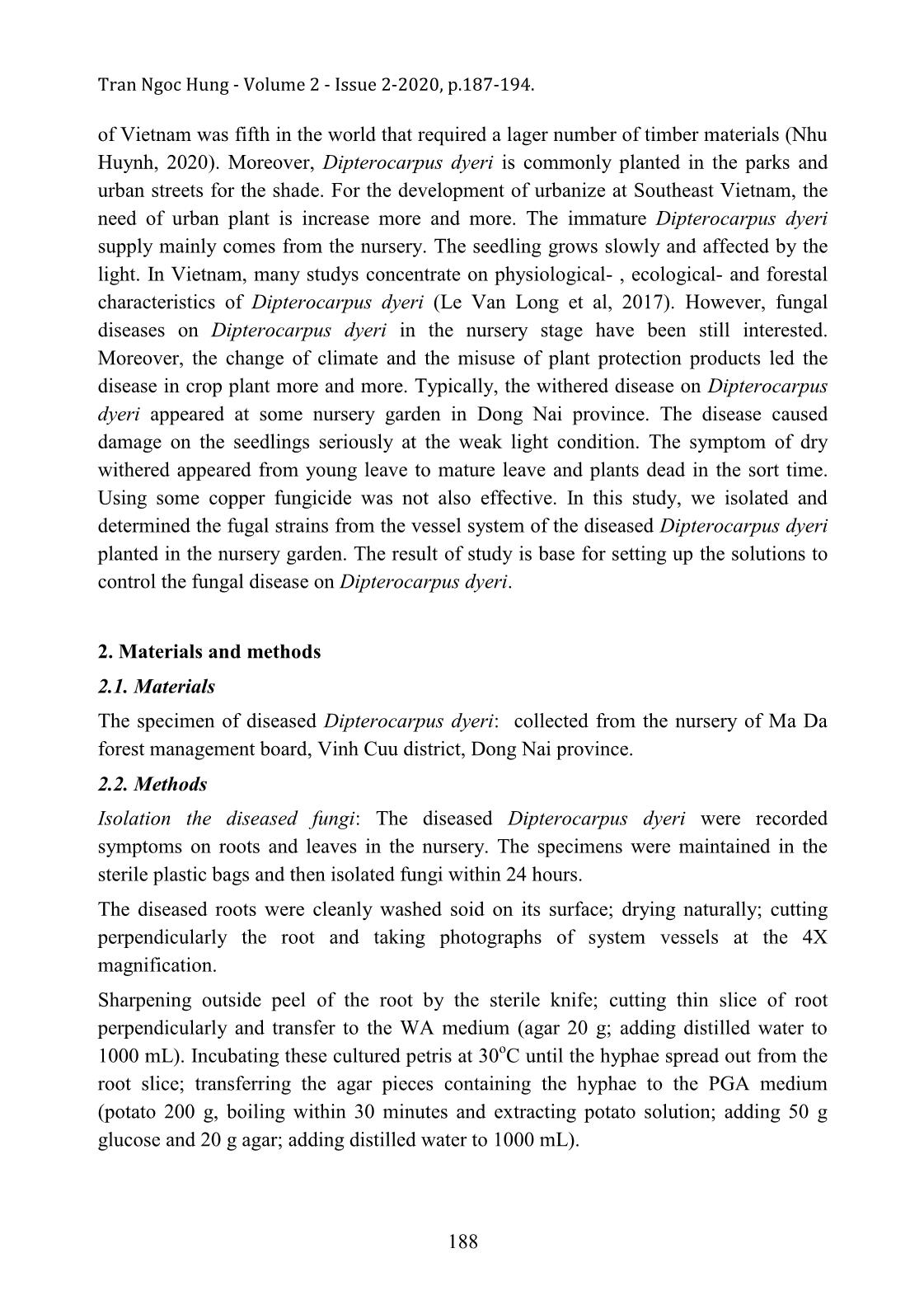
Recording symptoms of plants at the plantation and micro-characteristics of root at the lab
(figure 1); washing cleanly soil outside of the disease root of Dipterocarpus dyeri;
sharpening the peel of root; cutting thin layers of root and then putting them on WA
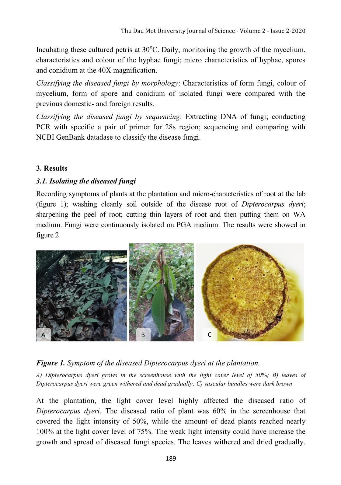
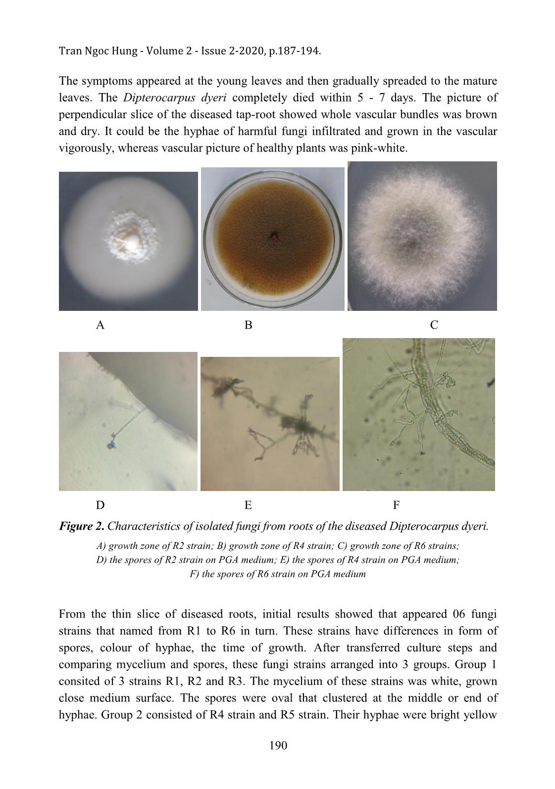
medium. Fungi were continuously isolated on PGA medium. The results were showed in
figure 2.
 A B C
Figure 1. Symptom of the diseased Dipterocarpus dyeri at the plantation.
A) Dipterocarpus dyeri grows in the screenhouse with the light cover level of 50%; B) leaves of
Dipterocarpus dyeri were green withered and dead gradually; C) vascular bundles were dark brown
At the plantation, the light cover level highly affected the diseased ratio of
Dipterocarpus dyeri. The diseased ratio of plant was 60% in the screenhouse that
covered the light intensity of 50%, while the amount of dead plants reached nearly
100% at the light cover level of 75%. The weak light intensity could have increase the
growth and spread of diseased fungi species. The leaves withered and dried gradually.
 189
Tran Ngoc Hung - Volume 2 - Issue 2-2020, p.187-194.
The symptoms appeared at the young leaves and then gradually spreaded to the mature
leaves. The Dipterocarpus dyeri completely died within 5 - 7 days. The picture of
perpendicular slice of the diseased tap-root showed whole vascular bundles was brown
and dry. It could be the hyphae of harmful fungi infiltrated and grown in the vascular
vigorously, whereas vascular picture of healthy plants was pink-white.
 A B C
 D E F
Figure 2. Characteristics of isolated fungi from roots of the diseased Dipterocarpus dyeri.
 A) growth zone of R2 strain; B) growth zone of R4 strain; C) growth zone of R6 strains;
 D) the spores of R2 strain on PGA medium; E) the spores of R4 strain on PGA medium;
 F) the spores of R6 strain on PGA medium
From the thin slice of diseased roots, initial results showed that appeared 06 fungi
strains that named from R1 to R6 in turn. These strains have differences in form of
spores, colour of hyphae, the time of growth. After transferred culture steps and
comparing mycelium and spores, these fungi strains arranged into 3 groups. Group 1
consited of 3 strains R1, R2 and R3. The mycelium of these strains was white, grown
close medium surface. The spores were oval that clustered at the middle or end of
hyphae. Group 2 consisted of R4 strain and R5 strain. Their hyphae were bright yellow
 190
 Thu Dau Mot University Journal of Science - Volume 2 - Issue 2-2020
or orange-yellow. Spores were spherical shape that formed from a bottle shaped conidia
and attached together into chain. Following the taxonomy described by Nguyen Huy
Van, R4 and R5 strain could belong to Aspergillus genus (Nguyen Huy Van and Bui
Xuan Dong, 2000) whereas, the hyphae of R6 strain were light grey. Its spores were
oval grey shape that formed separately at the end of mycelium. R2, R4 and R6 strain
were continuously classified by sequencing.
3.2. Classifying the diseased fungi by sequencing
The sequencing of 420 nucleotid of R2 ITS region (pair of primers were ITS1 and
ITS2):
CTTATTGATATGCTTAAGTTCAGCGGGTAGTCCTACCTGATCCGAGGTCAAC
CTTGTAAAAAAGAGTTGCACGTCATGCGCGATTTGCATGGCAGGGCGCCGG
CGGGGCTTCCGTAGCGAGAGGAGAGAACTTGCGTTCAGTACTGCGCTCGGA
GCCAGCCAGCCGGGCCCGCCACTCGCTTTCGGGGCCCTCCGCCAGGCGGAG
GAGCCCCAACACCAGCGCGCAACGGGGCGCGTGAGGGGGGAAATGACGCT
CGGACAGGCATGCCCGCCAGAATACTGGCGGGCGCAATGTGCGTTCAAAGA
TTCGATGGTTCGCTGAATTCTGCAATTCACATTACGTATCGCATTTCGCTGC
GTTCTTCATCGATGCCAGAGCCAAGAGATCCGTTGTTGAAAGTTTTAATGTA
TTTTTGTTTT
Comparing with NCBI GenBank datadase by BLAST SEARCH solfware showed R2
strain belong to Ophiostoma eucalyptigena with identity level of 100% (E-value =0).
The sequencing of 896 nucleotid of R4 ITS region (pair of primers were ITS1 and
ITS2):
CACACGGGCACGGGCACCCTGCCCAAGACGGGATTCTCACCCTCTCTGACG
GCCCGTTCCAGGGCACTTAGACAGGGGCCGCACCCGAAGCATCCTCTGCAA
ATTACAACGCGGACCCCGAAGGGGCCAGCTTTCAAATTTGAGCTCTTGCCG
CTTCACTCGCCGTTACTGAGGCAATCCCGGTTGGTTTCTTTTCCTCCGCTTAT
TGATATGCTTAAGTTCAGCGGGTATCCCTACCTGATCCGAGGTCAACCTGAG
AAAAATAAGGTTGGAGACGCCGGCTGGCGCCCGGCCGGCCCTAATCGAGCG
GGTGACAAAGCCCCATACGCTCGAGGACCGGACACGGTGCCGCCGCTGCCT
TTCGGGCCCGTCCCCCGGGGGGGACGACGACCCAACACACAAGCCGGGCTT
GAGGGCAGCAATGACGCTCGGACAGGCATGCCCCCCGGAATGCCAGGGGG
CGCAATGTGCGTTCAAAGACTCGATGATTCACTGAATTCTGCAATTCACATT
ACTTATCGCAGTTCGCTGCGTTCTTCATCGATGCCGGAACCAAGAGATCCAT
TGTTGAAAGTTTTGACTGATTTGTATTCAGGCTCAGACTGCATCACTCTCAG
GCATGAAGTTCGGTGGTCCCCGGCGGCTCGCCCCTGGGGGGCTCCCCGCCG
AAGCAACAGTGTTAGGTAGTCACGGGTGGGAGGTTGGGCGCCCGGAGGCA
GCCCGCACTCGGTAATGATCCTTCCGCAGGTTCACCTACGGAAACCTTGTTA
 191
Tran Ngoc Hung - Volume 2 - Issue 2-2020, p.187-194.
CGACTTTTACTTCCTCTAAATGACCGGGTTTGACCAGCTTTCCGGCTCGGGG
GGGTCGTTGCCAACCCTCCTGAGCCAGTCCGGAGGCCTCACCGAGCCATTC
AATCGGTAGTAGCGACGGGCGGT
Comparing with NCBI GenBank datadase by BLAST SEARCH solfware showed R4
strain belong to Aspergillus nidulans with identity level of 99% (E-value =0).
The sequencing of 904 nucleotid of R6 ITS region (pair of primers were ITS1 and
ITS2):
GGAGCTTTACAAAGGCTTGGTGTCCAACTGTACGGGGCTCTCACCCTCTCTG
GCGTCCCGTTCTAGGGAACTTGGAAGGCACCGCACCAAAAGCATCCTCTGC
AAATTACAACTCGGGCCTAGGGCCAGATTTCAAATTTGAGCTGTTGCCGCTT
CACTCGCCGTTACTGAGGCAATCCCTGTTGGTTTCTTTTCCTCCGCTTATTGA
TATGCTTAAGTTCAGCGGGTATTCCTACCTGATCCGAGGTCAACCTTTGGAA
AATTGGGGGGTTTTACGGCAAGAGTCCCTCCGGATCCCAGTGCGAGACGTA
AAGTTACTACGCAAAGGAGGCTCCGGGAGGGTCCGCCACTACCTTTGAGGG
CCTACATCAGCTGTAGGGCCCCAACACCAAGCAGAGCTTGAGGGTTGAAAT
GACGCTCGAACAGGCATGCCCGCCAGAATGCTGGCGGGCGCAATGTGCGTT
CAAAGATTCGATGATTCACTGAATTCTGCAATTCACATTACTTATCGCATTT
CGCTGCGTTCTTCATCGATGCCAGAACCAAGAGATCCGTTGTTAAAAGTTTT
GATTATTTGCTTGTACCACTCAGAAGAAACGTCGTTAAATCAGAGTTTGGTT
ATCCTCCGGCGGGCGCCGACCCGCCCGGAGGCGGGAGGCCGGGAGGGTCG
CGGAGACCCTACCCGCCGAAGCAACAGTTATAGGTATGTTCACAAAGGGTT
ATAGAGCGTAAACTCAGTAATGATCCCTCCGCTGGTTCACCAACGGAGACC
TTGTTACGACTTTTACTTCCTCTAAATGACCGAGTTTGGATAACTTTCCGACC
AGGGGGAAGAGTTGCCTCCTCCCTTAGATCAGTCCGAAAGCCTCACTGAGC
CATTCAATCGGTAGTAGCGACGGGCGGT
Comparing with NCBI GenBank datadase by BLAST SEARCH solfware showed R6 strain
belong to Collectotrichum gloeosporioides with identity level of 99% (E-value =0).
4. Discussion
The Dipterocarpaceae is typical plants in forest at SouthEast Vietnam. They were
popularly planted in parks or urban areas for shade. Timber of Dipterocarpaceae was
also important material for woodwork. For increasing demand, Dipterocarpus dyeri was
planted more and more. A high density of plants and fluctuant weather has appeared
many disease on plants that has not been reported ever, typically the withered disease on
Dipterocarpus dyeri plants that have just appeared recently at some plantations in Dong
Nai province. Following the initial recognition at the plantation, the immature plants
often withered seriously at the lack of light condition. This showed that the immature
 192
 Thu Dau Mot University Journal of Science - Volume 2 - Issue 2-2020
plants could be attacked by diseased fungi easily at high level of light cover in natural
forest and reduced regenerative ability of Dipterocarpus dyeri population in nature. The
picture of thin slice root showed its vessel was brown that was a sign of microbial
infiltration. On WA medium, it appeared many hyphae from the diseased root specimen
without bacteria.
Sequencing these fungi showed they belonged to Ophiostoma eucalyptigena,
Aspergillus nidulans and Collectotrichum gloeospoioides that identity level got 100%,
99% and 99%, respectively. Our result was relative resemble the others. Ophiostoma
eucalyptigena was refered by Pedro in the previous result. They caused the diseased
wither on large trees in Australia (Pedro, 2015). Collectotrichum gloeospoioides strain
was recognized to be cause of the anthracnose disease on many fruit trees such as
mango, papaya, grape, avocado, chili pepper Symptoms of the anthracnose disease
often commonly appear on ripe fruits and leaves (Erika et al, 2009 ; Kim et al, 2004 ;
Hyde et al, 2009). However, it has not reported that Collectotrichum gloeospoioides was
cause of wither disease on plants. Whereas Aspergillus nidulans has not recognized as
main cause of the disease on plants yet. Aspergillus genus was considered to be
saprophytic fungi. This strain could have infiltrated into the vascular when the plants
were injured. Classification of this fungi could provide more informations to control the
dieases on Diprterocarpus dyeri.
5. Conclution
On the wilted Dipterocarpus dyeri at the nursery, the study initially isolated 03 disease
fungi strains. Observing morphologic characteristic in conjunction with comparing gene
sequence showed that these fungi belonged to Ophiostoma eucalyptigena, Aspergillus
nidulans and Collectotrichum gloeospoioides with identity level of 100%, 99% and
99%, respectively. The results were a base for setting up solutions to control the wilt on
Dipterocarpus dyeri at the nursery.
 Acknowledgment
 We deeply thank Thu Dau Mot University and The Center of Practice and
 Research Thu Dau Mot for material facility support. We also thank Mrs Ha and
 Mr Trung at the nursery of Ma Da forest management board for immature
 plants and useful information.
References
Erika P., Martínez1, Juan C., Hío1, Jairo A., Osorio1 and María F. Torres, 2009. Identification
 of Colletotrichum species causing anthracnose on Tahiti lime, tree tomato and mango.
 Agronomía Colombiana. 27(2): 211-218.
 193
Tran Ngoc Hung - Volume 2 - Issue 2-2020, p.187-194.
Gavrias V., Timberlake W.E., Adam T.H., 2001. Aspergillus nidulans, Encyclopedia of Genetic.
 106-111. https://doi.org/10.1006/rwgn.2001.0082
Nhu Huynh, 2020. Overcoming many trades, 2019, exportation of wood and wood product gets
 a highest value in history. Available from: https://vietnambiz.vn/vuot-mat-nhieu-nganh-
 hang-nam-2019-xuat-khau-go-va-san-pham-go-dat-muc-cao-nhat-trong-lich-su-
 20200102112625027.htm. Accessed: 08/04/2020.
Hyde, K. D., Cai L., Cannon P.E., et al, 2009. Colletotrichum – names in current use. Fungal
 Diversity, 39: 147-182.
Kim K. K, Yoon J.B., Park H.G., Park E.W., Kim Y.H., 2004. Structural modifications and
 programmed cell death of chilli pepper fruits related to resistance responses to
 Colletotrichum gloeosporioides infection, Genetics and Resistance, 94: 1295-1304.
Le Van Long, Nguyen Minh Thanh, Le Van Cuong, Le Ba Toan, 2017. Some silvicutural
 characteristic of Dipterocarpus dyeri population in evergreen moist tropical forest at Tan
 Phu, Dong Nai provice. Journal of Sciencetific and Technology Forestry, 6: 42-50.
Lester W. Buger, Timothy E. Knight, Len Tesoriero, Phan Thuy Hien, 2009. Manual of disease
 diagnosis in Vietnam, Australian Centre for International Agricultural Research.
Pedro W.C. and Johannes Z.G., 2015. Ophiostoma eucalyptigena Barber & Crous, sp. nov,
 Fungal Planet, 331(34): 192-193.
Nguyen Minh Tam, 2016. Preservation gene sources of Dipterocarpus costatus and
 Dipterocarpus dyeri being threatened with extinction in Southeast Vietnam, Subject of
 Vietnam Academy of Science and Technology, Code VAST.BVMT.01/15-16.
Nguyen Huy Van and Bui Xuan Dong, 2000. Fungi in biotechnology, Pub. Science and
 Technology.
 194
File đính kèm:
 isolating_some_fungi_strains_from_diease_roots_of_dipterocar.pdf

