Dosimetric characteristics of 6 MV photons from TrueBeam STx medical linear accelerator: Simulation and experimental data
A TrueBeam STx is one of the most technologically advanced linear accelerators for
radiotherapy and radiosurgery. The Monte Carlo simulation widely used in many applications in
various fields such as nuclear physics, astrophysics, particle physics, and medicine. The
Geant4/GATE Monte Carlo toolkit is developed for the simulation in imaging diagnostics, nuclear
medicine, radiotherapy, and radiation biology to more accurately predict beam radiation dosimetry. In
this work, we present the simulation results of the dosimetric characteristics of a 6 MV photon beam
of TrueBeam STx medical LINAC using Monte Carlo Geant4/GATE. The percentage depth dose
(PDD), central axis depth dose (Profile) have been simulated and compared with those measured in a
water phantom for field sizes 10×10 cm2 via the gamma-index method. These results will permit to
check calculation data given by the treatment planning system.
Trang 1
Trang 2
Trang 3
Trang 4
Trang 5
Trang 6
Trang 7
Trang 8
Tóm tắt nội dung tài liệu: Dosimetric characteristics of 6 MV photons from TrueBeam STx medical linear accelerator: Simulation and experimental data
s of small fields and of ranges
with high dose gradients. The RAZOR has
sensitive volume and the radius cavity are 0.01
cm
3
 and 1.0 mm, respectively.
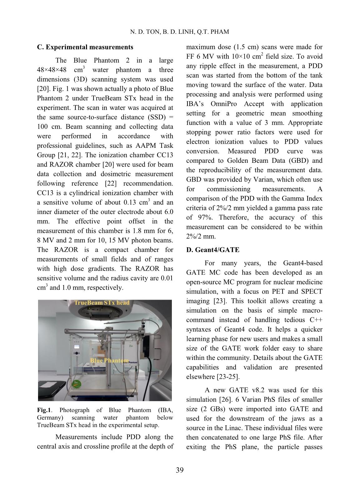
Fig.1. Photograph of Blue Phantom (IBA,
Germany) scanning water phantom below
TrueBeam STx head in the experimental setup.
Measurements include PDD along the
central axis and crossline profile at the depth of
maximum dose (1.5 cm) scans were made for
FF 6 MV with 10×10 cm
2
 field size. To avoid
any ripple effect in the measurement, a PDD
scan was started from the bottom of the tank
moving toward the surface of the water. Data
processing and analysis were performed using
IBA’s OmniPro Accept with application
setting for a geometric mean smoothing
function with a value of 3 mm. Appropriate
stopping power ratio factors were used for
electron ionization values to PDD values
conversion. Measured PDD curve was
compared to Golden Beam Data (GBD) and
the reproducibility of the measurement data.
GBD was provided by Varian, which often use
for commissioning measurements. A
comparison of the PDD with the Gamma Index
criteria of 2%/2 mm yielded a gamma pass rate
of 97%. Therefore, the accuracy of this
measurement can be considered to be within
2%/2 mm.
D. Geant4/GATE
For many years, the Geant4-based
GATE MC code has been developed as an
open-source MC program for nuclear medicine
simulation, with a focus on PET and SPECT
imaging [23]. This toolkit allows creating a
simulation on the basis of simple macro-
command instead of handling tedious C++
syntaxes of Geant4 code. It helps a quicker
learning phase for new users and makes a small
size of the GATE work folder easy to share
within the community. Details about the GATE
capabilities and validation are presented
elsewhere [23-25].
A new GATE v8.2 was used for this
simulation [26]. 6 Varian PhS files of smaller
size (2 GBs) were imported into GATE and
used for the downstream of the jaws as a
source in the Linac. These individual files were
then concatenated to one large PhS file. After
exiting the PhS plane, the particle passes
DOSIMETRIC CHARACTERISTICS OF 6 MV PHOTONS FROM TRUEBEAM STX
40
through the second collimator as the Y and X
jaws and MLC. Data for the material and
geometry of the Linac components were
obtained from the TrueBeam STx Monte Carlo
package [19].
Geant4 Electromagnetic physics package
3 (G4EmStandardPhysics_option3) was used
for precise dose calculations and particle-
matter interactions or radiation transport in the
simulation. G4EmStandardPhysics_option3
designed for any applications required higher
accuracy of electrons, hadrons, and ion
tracking without a magnetic field. The package
has been presented in Poon and Arce at al. for
radiotherapy application [27, 28] and
recommended in Varian documents [19]. The
range cuts for gamma, electron, and positron
are fixed to 0.1 mm in a water phantom, 1 mm
in the world volume, and 10 mm in TrueBeam
material volume, respectively.
A virtual water phantom with 30×30×30
cm
3
 volume was installed at an SSD equal to
100 cm as the measurement. It is used for the
MC estimation of the absorbed dose
distribution. Voxel size was set to 3×3×3 mm
2
for a field size of 10×10 cm
2
.
E. Gamma Index method
Data analysis was based on comparisons
between GATE simulations and measurements
using the Gamma Index method [29], which
became a ―gold standard‖ method for the
comparison in dose distribution [13]. This
method was conducted with a percentage dose
difference (ΔD) criteria and distance to
agreement (DTA) of of 1%/1mm and
2%/2mm. If the Gamma Index value is greater
than unity, it indicates a position where the
agreement between the measured and
simulated dose maps do not meet the
predefined criteria. Passing criteria were met if
the gamma index was no larger than 1. An
important feature of this method is that the
final assessment of the dose distribution
quality. For the regions of significant
disagreement, the Gamma Index value is
greater than unity that will be apparent relative.
The gamma pass rate was defined as a
quotient of the passing points and all points.
For the global Gamma Index passing criteria of
2%/2 mm, a good agreement, a high
agreement, and a reasonable agreement
between the measured and simulated dose
distribution were observed with over 99%,
95% and 90% of the points of PDD and cross-
plane profile, respectively.
III. RESULTS AND DISCUSSION
6 PhS files stored 3×10
8
 photon,
electron, and positron particles, which have
been simulated. Approximately 6×10
9
 particle
histories from 6 PhS files were performed such
that the statistical uncertainty in the dose for
the voxels inside the radiation field was less
than 0.2% at the depth of maximum water
phantom. The simulation results take into
account the electron and positron
contamination in the photon beam.
A. PDD curve
In this paper, the quantity PDD defined
as the quotient, expressed as a percentage, of
the absorbed dose at a predefined depth (dx) to
the absorbed maximum dose at a fixed
reference depth of d0 = 1.5 cm, along the
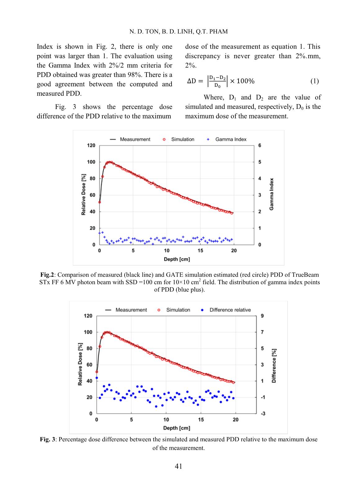
central axis of the beam. Fig. 2 shows the
comparison between measured and GATE
estimated PDD with SSD = 100 cm for a
10×10 cm
2
 field for FF 6 MV photon beam and
the Gamma Index distribution. The maximum
dose was detected at 1.5 cm of depth in both
measurement and simulation. The statistical
uncertainty of bins scoring PDD was between
0.02% to 0.04% and all bins scoring more than
50% of the maximum absorbed dose was
0.02% to 0.2%. The distribution of Gamma
N. D. TON, B. D. LINH, Q.T. PHAM
41
Index is shown in Fig. 2, there is only one
point was larger than 1. The evaluation using
the Gamma Index with 2%/2 mm criteria for
PDD obtained was greater than 98%. There is a
good agreement between the computed and
measured PDD.
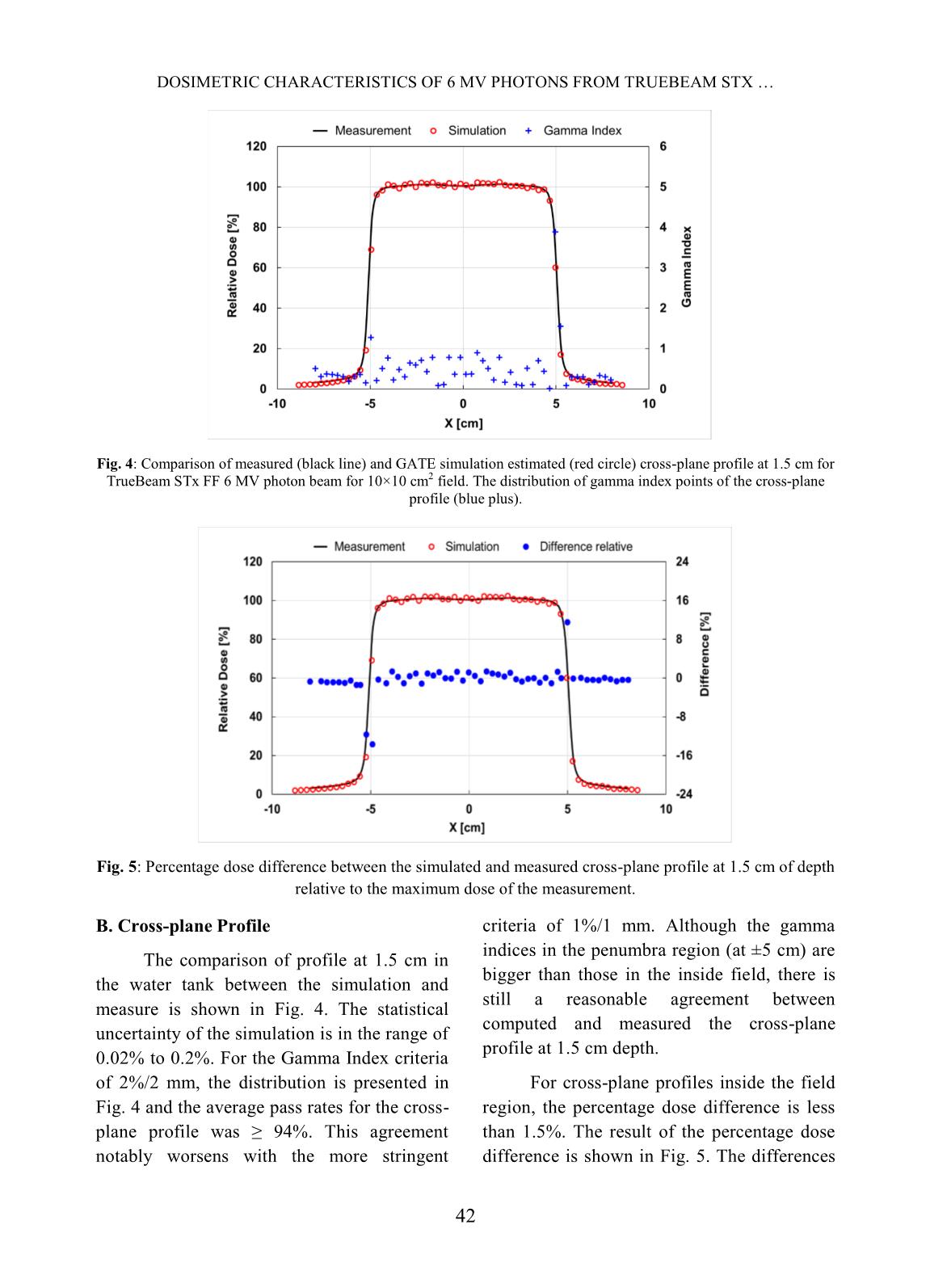
Fig. 3 shows the percentage dose
difference of the PDD relative to the maximum
dose of the measurement as equation 1. This
discrepancy is never greater than 2%.mm,
2%.
 |
| (1)
Where, D1 and D2 are the value of
simulated and measured, respectively, D0 is the
maximum dose of the measurement.
Fig.2: Comparison of measured (black line) and GATE simulation estimated (red circle) PDD of TrueBeam
STx FF 6 MV photon beam with SSD =100 cm for 10×10 cm
2
 field. The distribution of gamma index points
of PDD (blue plus).
Fig. 3: Percentage dose difference between the simulated and measured PDD relative to the maximum dose
of the measurement.
DOSIMETRIC CHARACTERISTICS OF 6 MV PHOTONS FROM TRUEBEAM STX
42
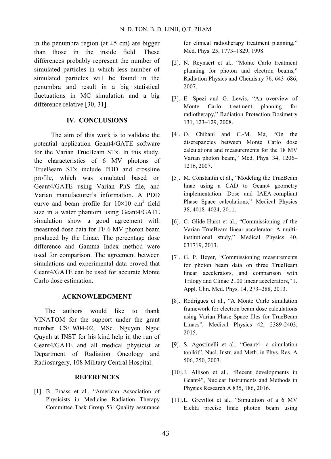
Fig. 4: Comparison of measured (black line) and GATE simulation estimated (red circle) cross-plane profile at 1.5 cm for
TrueBeam STx FF 6 MV photon beam for 10×10 cm2 field. The distribution of gamma index points of the cross-plane
profile (blue plus).
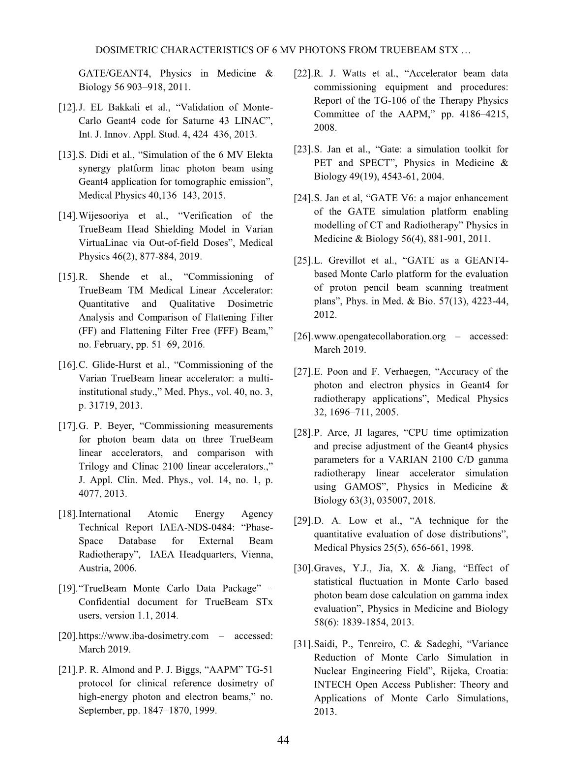
Fig. 5: Percentage dose difference between the simulated and measured cross-plane profile at 1.5 cm of depth
relative to the maximum dose of the measurement.
B. Cross-plane Profile
The comparison of profile at 1.5 cm in
the water tank between the simulation and
measure is shown in Fig. 4. The statistical
uncertainty of the simulation is in the range of
0.02% to 0.2%. For the Gamma Index criteria
of 2%/2 mm, the distribution is presented in
Fig. 4 and the average pass rates for the cross-
plane profile was ≥ 94%. This agreement
notably worsens with the more stringent
criteria of 1%/1 mm. Although the gamma
indices in the penumbra region (at ±5 cm) are
bigger than those in the inside field, there is
still a reasonable agreement between
computed and measured the cross-plane
profile at 1.5 cm depth.
For cross-plane profiles inside the field
region, the percentage dose difference is less
than 1.5%. The result of the percentage dose
difference is shown in Fig. 5. The differences
N. D. TON, B. D. LINH, Q.T. PHAM
43
in the penumbra region (at ±5 cm) are bigger
than those in the inside field. These
differences probably represent the number of
simulated particles in which less number of
simulated particles will be found in the
penumbra and result in a big statistical
fluctuations in MC simulation and a big
difference relative [30, 31].
IV. CONCLUSIONS
The aim of this work is to validate the
potential application Geant4/GATE software
for the Varian TrueBeam STx. In this study,
the characteristics of 6 MV photons of
TrueBeam STx include PDD and crossline
profile, which was simulated based on
Geant4/GATE using Varian PhS file, and
Varian manufacturer’s information. A PDD
curve and beam profile for 10×10 cm
2
 field
size in a water phantom using Geant4/GATE
simulation show a good agreement with
measured dose data for FF 6 MV photon beam
produced by the Linac. The percentage dose
difference and Gamma Index method were
used for comparison. The agreement between
simulations and experimental data proved that
Geant4/GATE can be used for accurate Monte
Carlo dose estimation.
ACKNOWLEDGMENT
The authors would like to thank
VINATOM for the support under the grant
number CS/19/04-02, MSc. Nguyen Ngoc
Quynh at INST for his kind help in the run of
Geant4/GATE and all medical physicist at
Department of Radiation Oncology and
Radiosurgery, 108 Military Central Hospital.
REFERENCES
[1]. B. Fraass et al., ―American Association of
Physicists in Medicine Radiation Therapy
Committee Task Group 53: Quality assurance
for clinical radiotherapy treatment planning,‖
Med. Phys. 25, 1773–1829, 1998.
[2]. N. Reynaert et al., ―Monte Carlo treatment
planning for photon and electron beams,‖
Radiation Physics and Chemistry 76, 643–686,
2007.
[3]. E. Spezi and G. Lewis, ―An overview of
Monte Carlo treatment planning for
radiotherapy,‖ Radiation Protection Dosimetry
131, 123–129, 2008.
[4]. O. Chibani and C.-M. Ma, ―On the
discrepancies between Monte Carlo dose
calculations and measurements for the 18 MV
Varian photon beam,‖ Med. Phys. 34, 1206–
1216, 2007.
[5]. M. Constantin et al., ―Modeling the TrueBeam
linac using a CAD to Geant4 geometry
implementation: Dose and IAEA-compliant
Phase Space calculations,‖ Medical Physics
38, 4018–4024, 2011.
[6]. C. Glide-Hurst et al., ―Commissioning of the
Varian TrueBeam linear accelerator: A multi-
institutional study,‖ Medical Physics 40,
031719, 2013.
[7]. G. P. Beyer, ―Commissioning measurements
for photon beam data on three TrueBeam
linear accelerators, and comparison with
Trilogy and Clinac 2100 linear accelerators,‖ J.
Appl. Clin. Med. Phys. 14, 273–288, 2013.
[8]. Rodrigues et al., ―A Monte Carlo simulation
framework for electron beam dose calculations
using Varian Phase Space files for TrueBeam
Linacs‖, Medical Physics 42, 2389-2403,
2015.
[9]. S. Agostinelli et al., ―Geant4—a simulation
toolkit‖, Nucl. Instr. and Meth. in Phys. Res. A
506, 250, 2003.
[10]. J. Allison et al., ―Recent developments in
Geant4‖, Nuclear Instruments and Methods in
Physics Research A 835, 186, 2016.
[11]. L. Grevillot et al., ―Simulation of a 6 MV
Elekta precise linac photon beam using
DOSIMETRIC CHARACTERISTICS OF 6 MV PHOTONS FROM TRUEBEAM STX
44
GATE/GEANT4, Physics in Medicine &
Biology 56 903–918, 2011.
[12]. J. EL Bakkali et al., ―Validation of Monte-
Carlo Geant4 code for Saturne 43 LINAC‖,
Int. J. Innov. Appl. Stud. 4, 424–436, 2013.
[13]. S. Didi et al., ―Simulation of the 6 MV Elekta
synergy platform linac photon beam using
Geant4 application for tomographic emission‖,
Medical Physics 40,136–143, 2015.
[14]. Wijesooriya et al., ―Verification of the
TrueBeam Head Shielding Model in Varian
VirtuaLinac via Out-of-field Doses‖, Medical
Physics 46(2), 877-884, 2019.
[15]. R. Shende et al., ―Commissioning of
TrueBeam TM Medical Linear Accelerator:
Quantitative and Qualitative Dosimetric
Analysis and Comparison of Flattening Filter
(FF) and Flattening Filter Free (FFF) Beam,‖
no. February, pp. 51–69, 2016.
[16]. C. Glide-Hurst et al., ―Commissioning of the
Varian TrueBeam linear accelerator: a multi-
institutional study.,‖ Med. Phys., vol. 40, no. 3,
p. 31719, 2013.
[17]. G. P. Beyer, ―Commissioning measurements
for photon beam data on three TrueBeam
linear accelerators, and comparison with
Trilogy and Clinac 2100 linear accelerators.,‖
J. Appl. Clin. Med. Phys., vol. 14, no. 1, p.
4077, 2013.
[18]. International Atomic Energy Agency
Technical Report IAEA-NDS-0484: ―Phase-
Space Database for External Beam
Radiotherapy‖, IAEA Headquarters, Vienna,
Austria, 2006.
[19]. ―TrueBeam Monte Carlo Data Package‖ –
Confidential document for TrueBeam STx
users, version 1.1, 2014.
[20]. https://www.iba-dosimetry.com – accessed:
March 2019.
[21]. P. R. Almond and P. J. Biggs, ―AAPM‖ TG-51
protocol for clinical reference dosimetry of
high-energy photon and electron beams,‖ no.
September, pp. 1847–1870, 1999.
[22]. R. J. Watts et al., ―Accelerator beam data
commissioning equipment and procedures:
Report of the TG-106 of the Therapy Physics
Committee of the AAPM,‖ pp. 4186–4215,
2008.
[23]. S. Jan et al., ―Gate: a simulation toolkit for
PET and SPECT‖, Physics in Medicine &
Biology 49(19), 4543-61, 2004.
[24]. S. Jan et al, ―GATE V6: a major enhancement
of the GATE simulation platform enabling
modelling of CT and Radiotherapy‖ Physics in
Medicine & Biology 56(4), 881-901, 2011.
[25]. L. Grevillot et al., ―GATE as a GEANT4-
based Monte Carlo platform for the evaluation
of proton pencil beam scanning treatment
plans‖, Phys. in Med. & Bio. 57(13), 4223-44,
2012.
[26]. www.opengatecollaboration.org – accessed:
March 2019.
[27]. E. Poon and F. Verhaegen, ―Accuracy of the
photon and electron physics in Geant4 for
radiotherapy applications‖, Medical Physics
32, 1696–711, 2005.
[28]. P. Arce, JI lagares, ―CPU time optimization
and precise adjustment of the Geant4 physics
parameters for a VARIAN 2100 C/D gamma
radiotherapy linear accelerator simulation
using GAMOS‖, Physics in Medicine &
Biology 63(3), 035007, 2018.
[29]. D. A. Low et al., ―A technique for the
quantitative evaluation of dose distributions‖,
Medical Physics 25(5), 656-661, 1998.
[30]. Graves, Y.J., Jia, X. & Jiang, ―Effect of
statistical fluctuation in Monte Carlo based
photon beam dose calculation on gamma index
evaluation‖, Physics in Medicine and Biology
58(6): 1839-1854, 2013.
[31]. Saidi, P., Tenreiro, C. & Sadeghi, ―Variance
Reduction of Monte Carlo Simulation in
Nuclear Engineering Field‖, Rijeka, Croatia:
INTECH Open Access Publisher: Theory and
Applications of Monte Carlo Simulations,
2013.
File đính kèm:
 dosimetric_characteristics_of_6_mv_photons_from_truebeam_stx.pdf

