Detection of ascaris suum in the livers of chickens infected naturally by the nested multiplex PCR assay
This study was conducted to detect Ascaris suum, Toxocara canis,
and Toxocara cati in naturally infected chicken livers, and then to
provide information related to ascarid infections in humans. Ninetyfour chicken liver samples collected at a fresh market were used for
this study. DNA was extracted from each minced liver sample (500
mg) by the alkaline lysis method using NaOH 50mM and Tris-HCl
(pH 8.0). The nested multiplex PCR assay using ascarid universal
primers was applied to amplify the ITS1 ribosomal RNA gene of A.
suum, Toxocara canis, and Toxocara cati in all the liver samples.
Then, species specific-primers were used to discriminate between A.
suum and Toxocara spp. The results showed that 2 of the 94 chicken
liver samples detected positive for the presence of A. suum DNA.
This study provided useful information and evidence about A. suum
infection in humans via exposure to contaminated soil or eating
raw/undercooked chicken livers in Vietnam.
Trang 1
Trang 2
Trang 3
Trang 4
Trang 5
Trang 6
Tóm tắt nội dung tài liệu: Detection of ascaris suum in the livers of chickens infected naturally by the nested multiplex PCR assay
turally infected
pigs, dogs, and cats, respectively, at local
abattoirs around Hanoi. They were identified by
morphology before performing the next steps.
The roundworms were then thoroughly washed
in saline solution and male worms were separated
and kept in 70% alcohol for DNA extraction.
Liver samples
Ninety-four chickens were purchased from a
fresh market in Trau Quy Town, Gia Lam
district, Hanoi, Vietnam in order to collect the
livers from naturally infected chickens. These
free-range chickens were reared freely and
derived from Hoa Binh, Bac Giang, and Hai
Detection of Ascaris suum in the Livers of chickens infected naturally by the nested multiplex PCR assay
608 Vietnam Journal of Agricultural Sciences
Duong provinces. The chickens then were
necropsied to inspect the macropathology (white
spot lesions) on the liver surfaces. Next, the
livers were collected for the detection of A. suum
by the nested multiplex PCR assay.
DNA extraction
The genomic DNA was extracted from adult
A. suum, T. canis, and T. cati worms using a
Blood- Animal- Plant preparation Kit (Jena
Bioscience, Germany) following the
manufacturer's instructions. DNA concentration
was measured by NanoDrop 2000
(ThermoFisher Scientific, US) to detect the
presence of DNA.
The alkaline-lysis method was employed for
extracting gDNA from the liver tissue. Five
hundred milligrams (500mg) of chicken liver
was homogenized in a 15mL tube provided by
BioMasher (Nippi, Tokyo, Japan), and 1.8 mL of
50mM NaOH was added. After boiling for 30
min, the samples were neutralized by 200μL of
1M Tris-HCl (pH 8.0). The mixture was vortexed
thoroughly and centrifuged at 14,000×g for
10min. The supernatant was transferred to a new
tube and stored at -20°C until analysis (Nguyen
et al., 2016).
Nested multiplex PCR assay
The nested multiplex PCR assay was applied
to amplify a region of the ITS1 ribosomal RNA
gene of A. suum, T. canis, and T. cati. The PCR
reaction was performed in the total volume of 25
μl containing 1μL of template DNA and the
following PCR mixture: 12.5μL Master mix 2X
(Phusa Biochem, Vietnam), 0.75μL of each
primer at concentrations of 0.3mM (Phusa
Biochem, Vietnam) and 10μL distilled water.
The thermal cycling profile was 94°C for 2min,
followed by 40 cycles with denaturation at 94°C
for 15s, primer annealing at 52°C for 30s, and
extending at 68°C for 1min, and a final
elongation step at 72oC for 7min. The first PCR
products were used as a template in the species-
specific nested multiplex PCR, in which multiple
primer sets were applied in the same tube with
0.75μL of each of species-specific forward and
reverse at concentrations 0.3mM. The PCR
conditions were similar to the first reaction, except
for primer annealing was at 57oC (Table 1) (Wang
et al., 2018).
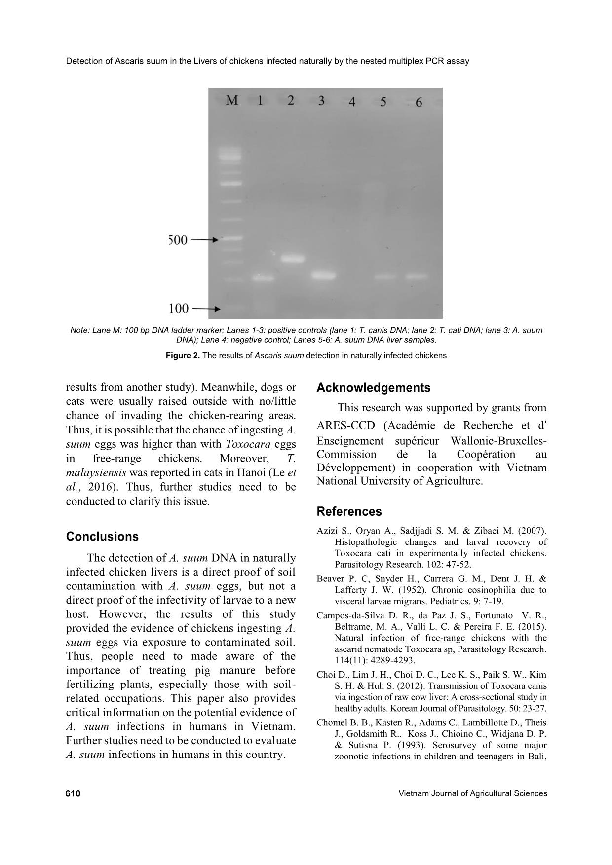
The amplification products were analyzed
using 1.0% agarose gel electrophoresis. The
amplification bands were visualized under UV
light. A positive reaction was observed when the
appearance of the specific sized bands of 208bp,
325bp, and 233bp corresponding with T. canis,
T. cati, and A. suum, respectively (Wang et al.,
2018).
Results and Discussion
Macroscopic inspection on the liver surfaces
Most of the collected livers were normal in
color and structure. However, 15 of the 94
chickens showed macropathological damage on
their livers. White spot lesions ranging from
Table 1. Primer sequences that were used for the nested PCR and nested multiplex PCR (Wang et al., 2018)
Primers Sequences
Ascarid specific universal
Primers (First PCR)
Ascarid
Forward: 5’-CGTCGGTAGCGATGAAAGGT-3’
Reverse: 5’-TTAGTTTCTTTTCCTCCGCT-3’
Species-specific primers
(second PCR)
T. canis
Forward: 5’-CTCGAGTCGACGAAGTATGTAC-3’
Reverse: 5’-AATTGGGCCGCCCATCATAC-3’
T. cati
Forward: 5’-GTAAGATCGTGGCACGCGTACGTA-3’
Reverse: 5’-TCTTTGATGTCAAGACTTCAGCGC-3’
A. suum
Forward: 5’-TCAACATTCATAGAGAATGGCATGT-3’
Reverse: 5’-TACATCATTATTGTCACGCTCATCT-3’
Nguyen Thị Hoang Yen et al. (2020)
https://vjas.vnua.edu.vn/ 609
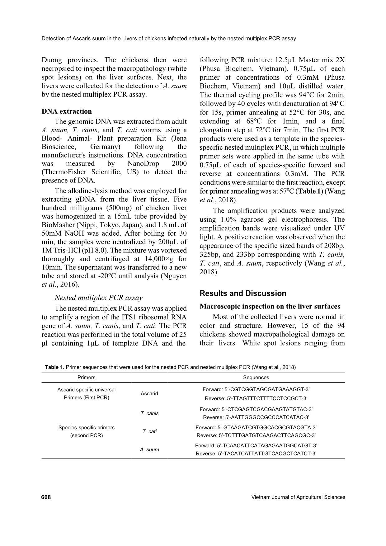
Figure 1. The white spot lesions on chicken livers infected naturally
1-10 in number and 0.1-0.5mm in diameter were
observed on both surfaces of the livers (Figure 1).
In addition, several different macropathological
features were observed on the liver surfaces, such
as damage caused by Marek’s disease. However,
those samples were excluded from this study.
The observation of white spot lesions on the
liver surfaces was one piece of critical evidence
of the migration of ascarid larvae through the
chicken liver (Taira et al., 2003; Azizi et al.,
2007; Yoshihara et al., 2008). However, the
white spot lesions were only recognized on day 7
in chickens with double or triple A. suum
infections (Yoshihara et al., 2008). White spot
lesions were not observed if the chickens were
not reinfected or in the cases of early infection
(Yoshihara et al., 2008).
In natural infection, chickens can ingest eggs
more frequently, leading to a higher chance of
larvae presenting in liver. Moreover, raw liver is
also a special cuisine in some countries such as
Japan and South Korea. This is one of the
transmission routes by which humans get ascarid
larva migrans syndrome (ascarid LMS) (Choi et
al., 2012). Thus, the liver samples were the target
of selection for this study.
Detection of A. suum in the chicken livers
Before applying the nested multiplex PCR
assay for the naturally infected liver samples,
positive control samples (ascarid adult-derived
DNA) were successfully amplified. Of the 94
livers collected, two of them were positive and
both of them showed amplification of a 233bp
band, which is specific for A. suum (Figure 2).
Meanwhile, Toxocara spp. DNA were not
detected in this study.
The presence and existence of larvae in the
liver depends on each agent. In the cases of T.
cati and A. suum, the larvae exist in the liver
around 14 days after the last experimental
infection (Azizi et al., 2007; Yoshihara et al.,
2008). For T. canis, after the larvae migrate to the
lungs, they migrate back to the liver and exist
there for ninety days (Okoshi & Usui, 1968;
Taira et al., 2003). The results of this study
showed that the two positive samples of A. suum
infection were not in livers showing white spot
lesions. Thus, the detection of A. suum herein
illustrated new infections of A. suum in those
naturally infected chickens. Investigation of
several free-range chicken farms showed that the
farmers often plant fruit trees such as pomelo
trees or litchi trees around and inside chicken-
rearing areas in order to provide shade for the
chickens, and they sometimes used pig manure to
fertilize these trees without treatment.
Additionally, chickens were reared in old
pigsties in some of the chicken farms (unpublished
Detection of Ascaris suum in the Livers of chickens infected naturally by the nested multiplex PCR assay
610 Vietnam Journal of Agricultural Sciences
Note: Lane M: 100 bp DNA ladder marker; Lanes 1-3: positive controls (lane 1: T. canis DNA; lane 2: T. cati DNA; lane 3: A. suum
DNA); Lane 4: negative control; Lanes 5-6: A. suum DNA liver samples.
Figure 2. The results of Ascaris suum detection in naturally infected chickens
results from another study). Meanwhile, dogs or
cats were usually raised outside with no/little
chance of invading the chicken-rearing areas.
Thus, it is possible that the chance of ingesting A.
suum eggs was higher than with Toxocara eggs
in free-range chickens. Moreover, T.
malaysiensis was reported in cats in Hanoi (Le et
al., 2016). Thus, further studies need to be
conducted to clarify this issue.
Conclusions
The detection of A. suum DNA in naturally
infected chicken livers is a direct proof of soil
contamination with A. suum eggs, but not a
direct proof of the infectivity of larvae to a new
host. However, the results of this study
provided the evidence of chickens ingesting A.
suum eggs via exposure to contaminated soil.
Thus, people need to made aware of the
importance of treating pig manure before
fertilizing plants, especially those with soil-
related occupations. This paper also provides
critical information on the potential evidence of
A. suum infections in humans in Vietnam.
Further studies need to be conducted to evaluate
A. suum infections in humans in this country.
Acknowledgements
This research was supported by grants from
ARES-CCD (Académie de Recherche et d’
Enseignement supérieur Wallonie-Bruxelles-
Commission de la Coopération au
Développement) in cooperation with Vietnam
National University of Agriculture.
References
Azizi S., Oryan A., Sadjjadi S. M. & Zibaei M. (2007).
Histopathologic changes and larval recovery of
Toxocara cati in experimentally infected chickens.
Parasitology Research. 102: 47-52.
Beaver P. C, Snyder H., Carrera G. M., Dent J. H. &
Lafferty J. W. (1952). Chronic eosinophilia due to
visceral larvae migrans. Pediatrics. 9: 7-19.
Campos-da-Silva D. R., da Paz J. S., Fortunato V. R.,
Beltrame, M. A., Valli L. C. & Pereira F. E. (2015).
Natural infection of free-range chickens with the
ascarid nematode Toxocara sp, Parasitology Research.
114(11): 4289-4293.
Choi D., Lim J. H., Choi D. C., Lee K. S., Paik S. W., Kim
S. H. & Huh S. (2012). Transmission of Toxocara canis
via ingestion of raw cow liver: A cross-sectional study in
healthy adults. Korean Journal of Parasitology. 50: 23-27.
Chomel B. B., Kasten R., Adams C., Lambillotte D., Theis
J., Goldsmith R., Koss J., Chioino C., Widjana D. P.
& Sutisna P. (1993). Serosurvey of some major
zoonotic infections in children and teenagers in Bali,
Nguyen Thị Hoang Yen et al. (2020)
https://vjas.vnua.edu.vn/ 611
Indonesia. Southeast Asian Journal of Tropical
Medicine Public Health. 24: 321-326.
Do T. D., Tran T. D., Nguyen T. H., Hoang Q. V., Do T. T.
T. & Nguyen T. L. A. (2016). Current status of
Toxocara spp. infection in humans in some study sites
of Hanoi and Hung Yen, 2014-2015. Journal of
Malaria and Parasitic Disease Control. 6(102): 10-16
(in Vietnamese).
Hoenigl M., Valentine T., Zollner-schwetz I., Salzer H. J.
F., Raggam R. B., Strenger V. & Krause R. (2010).
Pulmonary ascariasis: Two cases in Austria ANS
review of the literature. Wiener klinische
Wochenschrift. 122: 94-96.
Inatomi Y., Murakami T., Tokunaga M., Ishiwata K., Nawa
Y. & Uchino M. (1999). Encephalopathy caused by
visceral larva migrans due to Ascaris suum. Journal of
the Neurological Science. 164: 195-196.
Izumikawa K., Kohno Y., Izumikawa K., Hara H., Hayashi
H., Maruyama H. & Kohno S. (2011). Eosinophilic
pneumonia due to visceral larva migrans possibly
caused by Ascaris suum: A case report and review of
recent literatures. Japanese Journal of Infectious
Diseases. 64: 428-432.
Lamberton P. H. L. & Jourdan P. M. (2015). Human
Ascariasis: Diagnostics update. Current Tropical
Medicine Reports. 2: 189-200.
Magnaval J. F., Michault A., Calon N. & Charlet J. P.
(1994). Epidemiology of human toxocariasis in La
Reunion. Transactions of the Royal Society of Tropical
Medicine and Hygiene. 88: 531-533.
Maruyama H., Nawa Y., Noda S., Mimori T. & Choi W. Y.
(1996). An outbreak of visceral larva migrans due to
Ascaris suum in Kyushu, Japan. The Lancet. 347:
1766-1767.
Miller L. A., Colby K., Manning S. E., Hoenig D., McEvoy
E., Montgomery S. & Sears S. (2015). Ascariasis in
humans and pigs on small-scale farms, maine, USA,
2010-2013. Emerging Infectious Diseases. 21: 332-334.
Le T. H., Nguyen T. L. A., Nguyen T. K., Nguyen T. B. N.,
Do T. T. T. & Gasser R. B. (2016). Toxocara
malaysiensis infection in domestic cats in Vietnam – A
emerging zoonotic issue? Infection, Genetic and
Evolution. 37: 94-98.
Nguyen Y. T. H., Wang Z. Z., Maruyama H., Horii Y.,
Nonaka N. & Yoshida A. (2016). Evaluation of real-
time PCR assay for the detection of Ascaris suum
contamination in meat and organ meats. Journal of
Food Safety. 37(2): 1-6.
Okoshi S. & Ushui M. (1968). Experimental Studies on
Toxascaris Leonina. VI. Experimental Infection of
Mice, Chicken and Earthworms with Toxascaris
leonina, Toxocara canis and Toxocara cati. Japanese
Journal of Veterinary Medicine Science. 30: 151-166.
Pinelli E., Herremans T., Harms M. G., Hoek D. &
Kortbeek L. M. (2011). Toxocara and Ascaris
seropositivity among patients suspected of visceral and
ocular larva migrans in the netherlands: Trends from
1998 to 2009. European Journal of Clinical
Microbiology & Infectious Diseases: Official
Publication of the European Society of Clinical
Microbiology. 30: 873-879.
Umehara F., Ookatsu H., Hayashi D., Uchida A., Douchi
Y., Kawabata H. & Osame M. (2006). MRI studies of
spinal visceral larva migrans syndrome. Journal of
Neurological Science. 249: 7-12.
Sakakibara A., Basa K., Niwa S., Yagi T., Wakayama H.,
Yoshida K. & Kimura E. (2002). Visceral larva
migrans due to Ascaris suum which presented with
eosinophilic pneumonia and multiple intra-hepatic
lesions with severe eosinophil infiltration- outbreak in
a Japanese area other than Kuyshu. Internal Medicine.
41: 574-579.
Taira K., Permin A. & Kapel C. M. O. (2003).
Establishment and migration pattern of Toxocara canis
larvae in chickens. Parasitology Research. 90: 521-
523.
Thompson D. E., Bundy D. A. P., Cooper E. S. & Schantz
P. M. (1986). Epidemiological characteristics of
Toxocara canis infection of children in a Carribean
community. Bull WHO. 64: 283-290.
Tokojima M., Ashitani J. & Nakazato M. (2004). A case of
eosinophilic pneumonia caused by visceral larva
migrans due to Ascaris suum. The Japanese
Association for Infectious Diseases. 78: 1036-1040.
Tran T. D. (2014). Actual situation study, some factors has
risk to infect larva of Toxocara canis in human and
effect of treatment by Albendazole at 2 commune of
An Nhon District, Binh Dinh Province (2011-2012).
PhD thesis (in Vietnamese).
Vu T. K. V., Tran M. T. & Dang T. T. S. (2007). A survey
of manure management on pig farms in Northern
Viennam. Livestock Science. 112: 288-297.
Wang Z. Z., Shibata M., Nguyen Y. T. H., Hayata Y.,
Nonaka N., Maruyama H. & Yoshida A. (2018).
Development of nested multiplex polymerase chain
reaction (PCR) assay for the detection of Toxocara
canis, Toxocara cati and Ascaris suum contamination
in meat and organ meats. Parasitology International.
67: 622-628.
Yoshihara S., Hattori J., Nishizono K., Kawamura A.,
Shimozaki K., Nishida Y. & Hirayama N. (2008).
Hepatic lesions caused by migrating larvae of Ascaris
suum in chickens. Journal of Veterinary Medicine
Science. 70: 1129-1131.
File đính kèm:
 detection_of_ascaris_suum_in_the_livers_of_chickens_infected.pdf

