Application of morphological image processing to remove hair noises while enhancing patterns of near-Infrared human vein images
Recently, the innovative vein finder system has been studied
extensively and has many practical uses in healthcare and security. Developing a
better vein finder system often relies on image processing procedures which help to
enhance the vein images. Conventional image processing procedures as median
filtering and adaptive histogram equalization have shown benefit in enhancing vein
patterns. However, in some cases when there are hairs present in the images, most
of these procedures are less effective in removing noises from hairs. In this work,
we present a new approach employed additional morphological image processing
procedures to efficiently remove hair noises. We have successfully constructed a
vein finder device to acquire vein images and demonstrate the advantage of our
approach. Effects of the size and shape of the structural element in different
morphological image processing steps were studied and optimized to achieve the
best enhancement effect. Our approach can be applied widely to other vein finder
systems and enhance vein images from various parts of the human body.
Trang 1
Trang 2
Trang 3
Trang 4
Trang 5
Trang 6
Trang 7
Tóm tắt nội dung tài liệu: Application of morphological image processing to remove hair noises while enhancing patterns of near-Infrared human vein images
nder NIR illumination, veins appear as dark lines while skin and fat tissues
appear as lighter region, hence they are distinguishable. Developing a vein finder
system using NIR techniques [7] often faces a challenge, which is the low contrast
images that are generally present by light scattering. Enhancement of the contrast
of vein images is possible by image processing. Recent years, typical image
processing methods to enhance vein images have been used by many groups, both
Vietnamese [2, 5, 8] and international [3, 9], employed common procedures
including: application of median filtering, local histogram equalization following
by threshold filtering and sometimes Gaussian filtering.
However, in some cases, hairs on hands or legs of a human can be dense and
form a noise in the NIR vein images. The black hair acts just like a black body and
can absorb the full spectrum of light. Hence it is almost impossible to separate hair
noises/artifacts from vein patterns in NIR vein image by changing the optical
Journal of Military Science and Technology, Special Issue, No.72A, 5 - 2021 69
Biomedical Physics
configurations of the vein finder system. The hair noises also turn up as continuous
and thin lines like vein lines. Most of the conventional image processing
procedures appear to be less effective in removing noises from hairs. In this work,
we propose new image processing procedures using additional morphological
image processing steps to minimize (or to remove completely in some cases) noise
effect of hairs, while preserving the continuous vein lines. To demonstrate the
advantage of our approach, we construct a near-infrared vein imaging device to
acquire vein images, and apply and compare the conventional image processing
procedures versus ours.
2. MATERIALS AND METHODS
2.1. A near-infrared vein imaging device
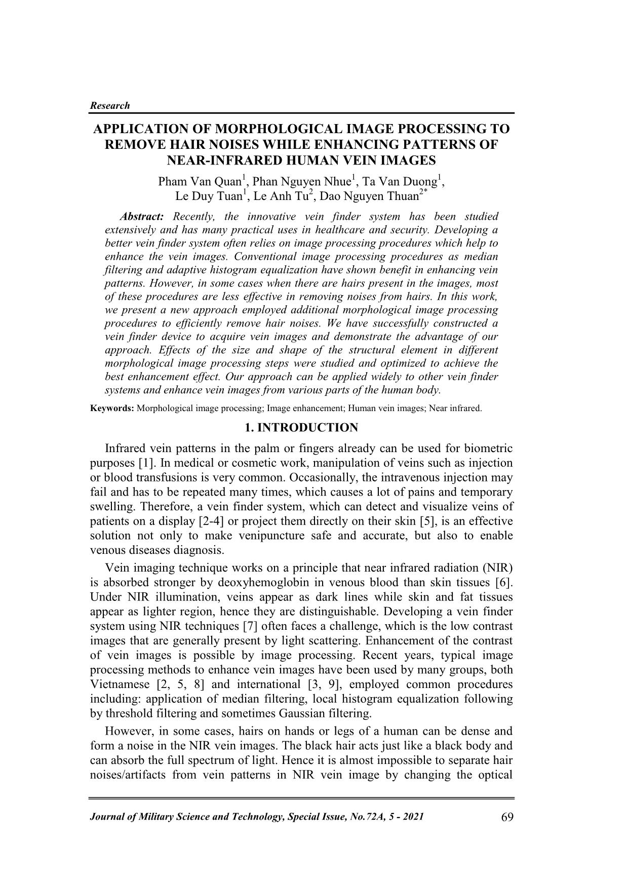
Figure 1. Diagram of the working principles of our near-infrared vein imaging
device (left side) and picture of the real device (right side).
We have constructed a near-infrared vein imaging device, which consisted of a
light source (850-nm or 950-nm LEDs), a camera to capture the vein images, a
computer Raspberry Pi 4 (model B) to process the digital image and a screen to
display output vein images (figure 1). The image processing program is developed
in Python language with open source library OpenCV and can be loaded and run
on the Raspberry Pi 4.
2.2. Conventional Image Processing Procedures
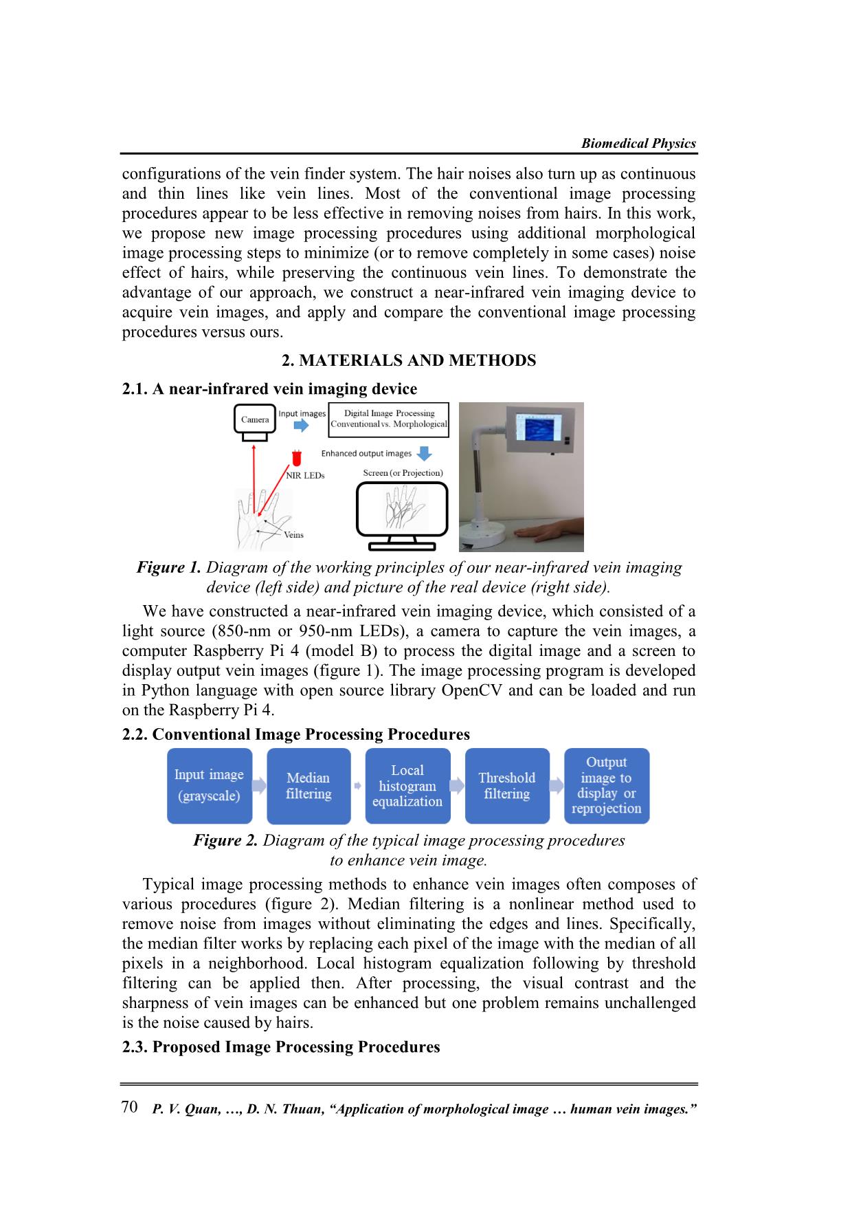
Figure 2. Diagram of the typical image processing procedures
to enhance vein image.
Typical image processing methods to enhance vein images often composes of
various procedures (figure 2). Median filtering is a nonlinear method used to
remove noise from images without eliminating the edges and lines. Specifically,
the median filter works by replacing each pixel of the image with the median of all
pixels in a neighborhood. Local histogram equalization following by threshold
filtering can be applied then. After processing, the visual contrast and the
sharpness of vein images can be enhanced but one problem remains unchallenged
is the noise caused by hairs.
2.3. Proposed Image Processing Procedures
70 P. V. Quan, , D. N. Thuan, “Application of morphological image human vein images.”
Research
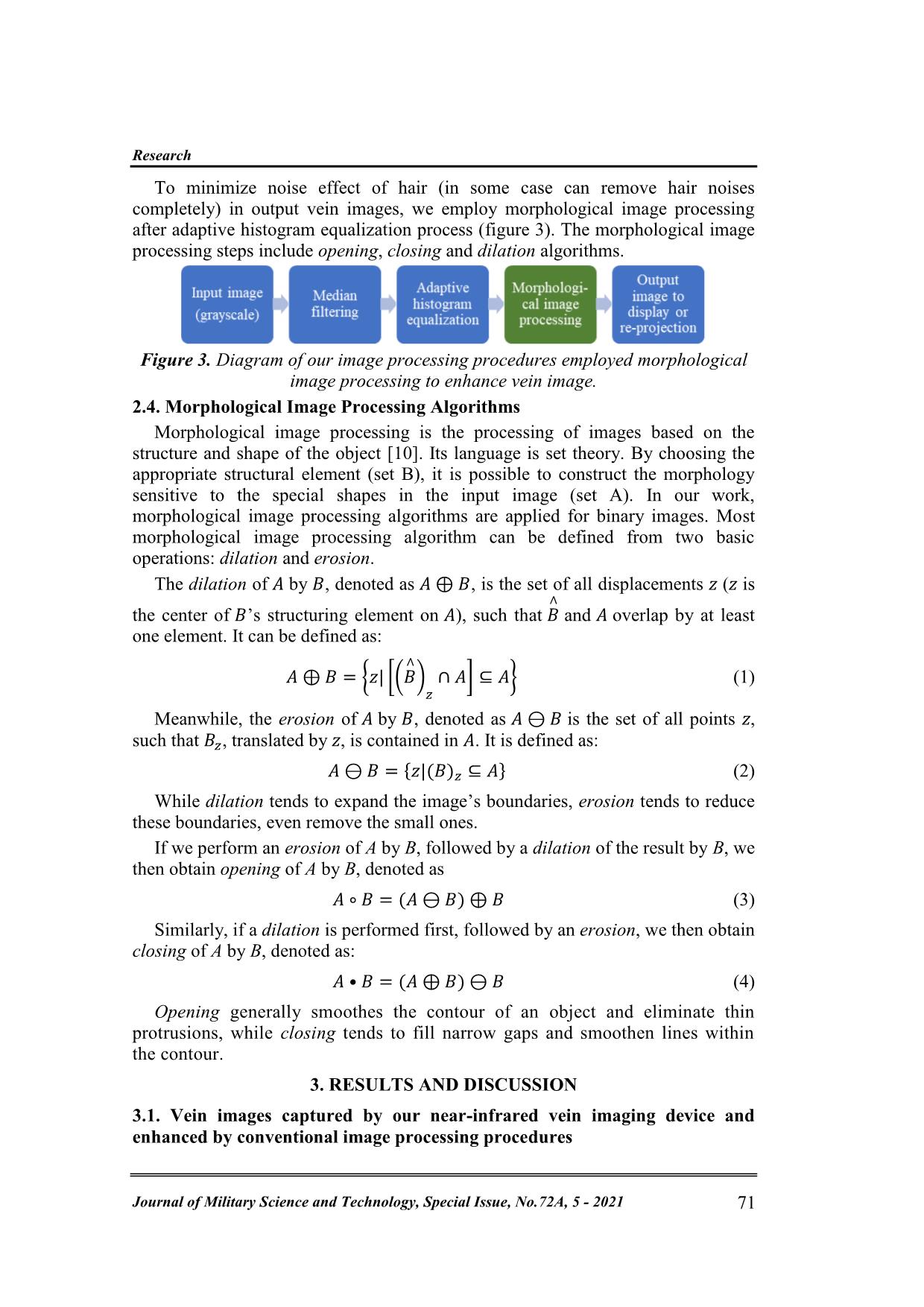
To minimize noise effect of hair (in some case can remove hair noises
completely) in output vein images, we employ morphological image processing
after adaptive histogram equalization process (figure 3). The morphological image
processing steps include opening, closing and dilation algorithms.
Figure 3. Diagram of our image processing procedures employed morphological
image processing to enhance vein image.
2.4. Morphological Image Processing Algorithms
Morphological image processing is the processing of images based on the
structure and shape of the object [10]. Its language is set theory. By choosing the
appropriate structural element (set B), it is possible to construct the morphology
sensitive to the special shapes in the input image (set A). In our work,
morphological image processing algorithms are applied for binary images. Most
morphological image processing algorithm can be defined from two basic
operations: dilation and erosion.
The dilation of by , denoted as , is the set of all displacements z (z is
the center of ’s structuring element on ), such that and overlap by at least
one element. It can be defined as:
{ [( ) ] } (1)
Meanwhile, the erosion of by , denoted as is the set of all points z,
such that , translated by z, is contained in . It is defined as:
{ } (2)
While dilation tends to expand the image’s boundaries, erosion tends to reduce
these boundaries, even remove the small ones.
If we perform an erosion of A by B, followed by a dilation of the result by B, we
then obtain opening of A by B, denoted as
(3)
Similarly, if a dilation is performed first, followed by an erosion, we then obtain
closing of A by B, denoted as:
(4)
Opening generally smoothes the contour of an object and eliminate thin
protrusions, while closing tends to fill narrow gaps and smoothen lines within
the contour.
3. RESULTS AND DISCUSSION
3.1. Vein images captured by our near-infrared vein imaging device and
enhanced by conventional image processing procedures
Journal of Military Science and Technology, Special Issue, No.72A, 5 - 2021 71
Biomedical Physics
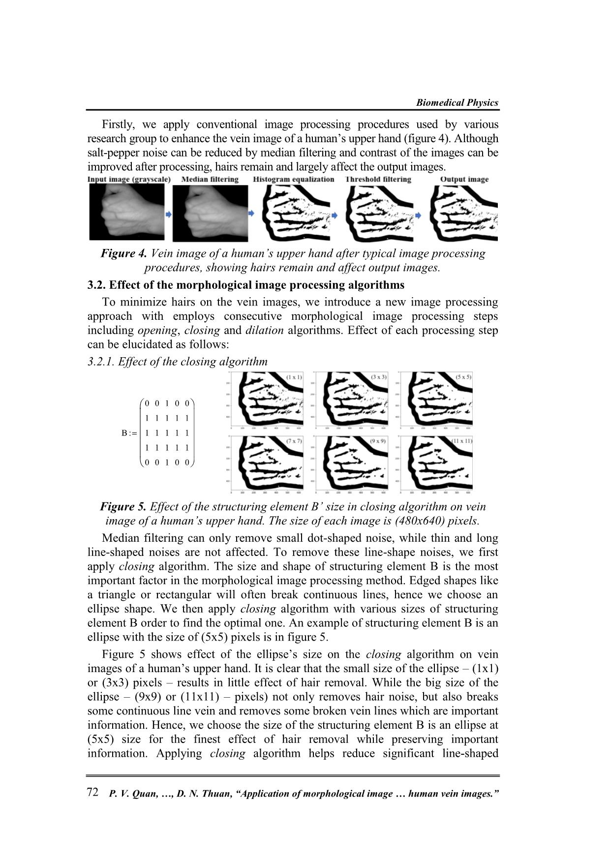
Firstly, we apply conventional image processing procedures used by various
research group to enhance the vein image of a human’s upper hand (figure 4). Although
salt-pepper noise can be reduced by median filtering and contrast of the images can be
improved after processing, hairs remain and largely affect the output images.
Figure 4. Vein image of a human’s upper hand after typical image processing
procedures, showing hairs remain and affect output images.
3.2. Effect of the morphological image processing algorithms
To minimize hairs on the vein images, we introduce a new image processing
approach with employs consecutive morphological image processing steps
including opening, closing and dilation algorithms. Effect of each processing step
can be elucidated as follows:
3.2.1. Effect of the closing algorithm
0 0 1 0 0
1 1 1 1 1
B 1 1 1 1 1
1 1 1 1 1
0 0 1 0 0
Figure 5. Effect of the structuring element B’ size in closing algorithm on vein
image of a human’s upper hand. The size of each image is (480x640) pixels.
Median filtering can only remove small dot-shaped noise, while thin and long
line-shaped noises are not affected. To remove these line-shape noises, we first
apply closing algorithm. The size and shape of structuring element B is the most
important factor in the morphological image processing method. Edged shapes like
a triangle or rectangular will often break continuous lines, hence we choose an
ellipse shape. We then apply closing algorithm with various sizes of structuring
element B order to find the optimal one. An example of structuring element B is an
ellipse with the size of (5x5) pixels is in figure 5.
Figure 5 shows effect of the ellipse’s size on the closing algorithm on vein
images of a human’s upper hand. It is clear that the small size of the ellipse – (1x1)
or (3x3) pixels – results in little effect of hair removal. While the big size of the
ellipse – (9x9) or (11x11) – pixels) not only removes hair noise, but also breaks
some continuous line vein and removes some broken vein lines which are important
information. Hence, we choose the size of the structuring element B is an ellipse at
(5x5) size for the finest effect of hair removal while preserving important
information. Applying closing algorithm helps reduce significant line-shaped
72 P. V. Quan, , D. N. Thuan, “Application of morphological image human vein images.”
Research
noises, however, it also cut some of the thin-lined veins. A simple closing algorithm
can not solve the problem and we have to use other following algorithms.
3.2.2. Effect of the opening algorithm
Using the same principle for testing resulted image with different sizes of
structuring element B, we derive the optimal size of the ellipse for the following
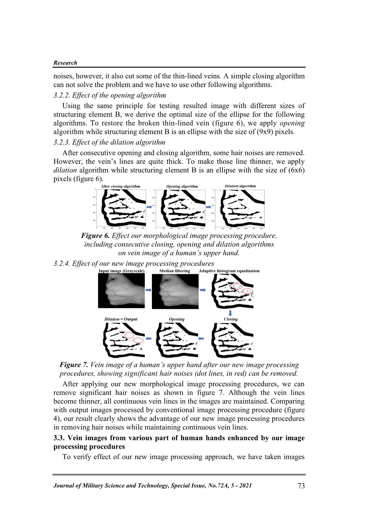
algorithms. To restore the broken thin-lined vein (figure 6), we apply opening
algorithm while structuring element B is an ellipse with the size of (9x9) pixels.
3.2.3. Effect of the dilation algorithm
After consecutive opening and closing algorithm, some hair noises are removed.
However, the vein’s lines are quite thick. To make those line thinner, we apply
dilation algorithm while structuring element B is an ellipse with the size of (6x6)
pixels (figure 6).
Figure 6. Effect our morphological image processing procedure,
including consecutive closing, opening and dilation algorithms
on vein image of a human’s upper hand.
3.2.4. Effect of our new image processing procedures
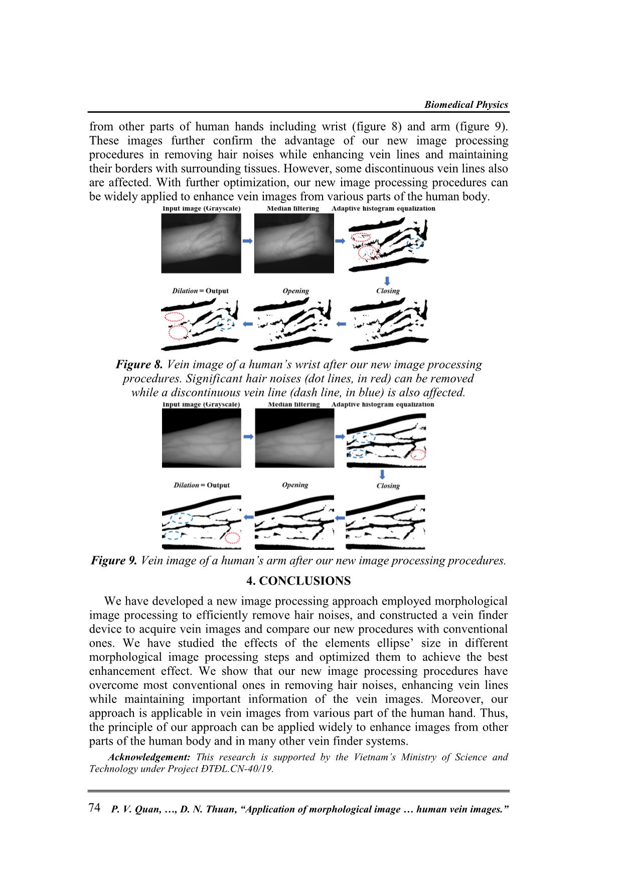
Figure 7. Vein image of a human’s upper hand after our new image processing
procedures, showing significant hair noises (dot lines, in red) can be removed.
After applying our new morphological image processing procedures, we can
remove significant hair noises as shown in figure 7. Although the vein lines
become thinner, all continuous vein lines in the images are maintained. Comparing
with output images processed by conventional image processing procedure (figure
4), our result clearly shows the advantage of our new image processing procedures
in removing hair noises while maintaining continuous vein lines.
3.3. Vein images from various part of human hands enhanced by our image
processing procedures
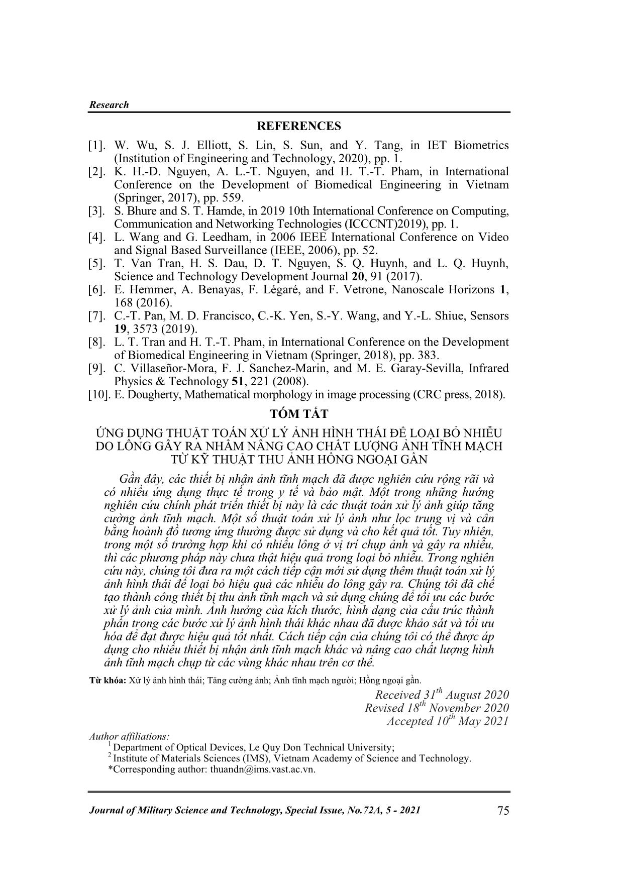
To verify effect of our new image processing approach, we have taken images
Journal of Military Science and Technology, Special Issue, No.72A, 5 - 2021 73
Biomedical Physics
from other parts of human hands including wrist (figure 8) and arm (figure 9).
These images further confirm the advantage of our new image processing
procedures in removing hair noises while enhancing vein lines and maintaining
their borders with surrounding tissues. However, some discontinuous vein lines also
are affected. With further optimization, our new image processing procedures can
be widely applied to enhance vein images from various parts of the human body.
Figure 8. Vein image of a human’s wrist after our new image processing
procedures. Significant hair noises (dot lines, in red) can be removed
while a discontinuous vein line (dash line, in blue) is also affected.
Figure 9. Vein image of a human’s arm after our new image processing procedures.
4. CONCLUSIONS
We have developed a new image processing approach employed morphological
image processing to efficiently remove hair noises, and constructed a vein finder
device to acquire vein images and compare our new procedures with conventional
ones. We have studied the effects of the elements ellipse’ size in different
morphological image processing steps and optimized them to achieve the best
enhancement effect. We show that our new image processing procedures have
overcome most conventional ones in removing hair noises, enhancing vein lines
while maintaining important information of the vein images. Moreover, our
approach is applicable in vein images from various part of the human hand. Thus,
the principle of our approach can be applied widely to enhance images from other
parts of the human body and in many other vein finder systems.
Acknowledgement: This research is supported by the Vietnam’s Ministry of Science and
Technology under Project ĐTĐL.CN-40/19.
74 P. V. Quan, , D. N. Thuan, “Application of morphological image human vein images.”
Research
REFERENCES
[1]. W. Wu, S. J. Elliott, S. Lin, S. Sun, and Y. Tang, in IET Biometrics
(Institution of Engineering and Technology, 2020), pp. 1.
[2]. K. H.-D. Nguyen, A. L.-T. Nguyen, and H. T.-T. Pham, in International
Conference on the Development of Biomedical Engineering in Vietnam
(Springer, 2017), pp. 559.
[3]. S. Bhure and S. T. Hamde, in 2019 10th International Conference on Computing,
Communication and Networking Technologies (ICCCNT)2019), pp. 1.
[4]. L. Wang and G. Leedham, in 2006 IEEE International Conference on Video
and Signal Based Surveillance (IEEE, 2006), pp. 52.
[5]. T. Van Tran, H. S. Dau, D. T. Nguyen, S. Q. Huynh, and L. Q. Huynh,
Science and Technology Development Journal 20, 91 (2017).
[6]. E. Hemmer, A. Benayas, F. Légaré, and F. Vetrone, Nanoscale Horizons 1,
168 (2016).
[7]. C.-T. Pan, M. D. Francisco, C.-K. Yen, S.-Y. Wang, and Y.-L. Shiue, Sensors
19, 3573 (2019).
[8]. L. T. Tran and H. T.-T. Pham, in International Conference on the Development
of Biomedical Engineering in Vietnam (Springer, 2018), pp. 383.
[9]. C. Villaseñor-Mora, F. J. Sanchez-Marin, and M. E. Garay-Sevilla, Infrared
Physics & Technology 51, 221 (2008).
[10]. E. Dougherty, Mathematical morphology in image processing (CRC press, 2018).
TÓM TẮT
ỨNG DỤNG THUẬT TOÁN XỬ LÝ ẢNH HÌNH THÁI ĐỂ LOẠI BỎ NHIỄU
DO LÔNG GÂY RA NHẰM NÂNG CAO CHẤT LƯỢNG ẢNH TĨNH MẠCH
TỪ KỸ THUẬT THU ẢNH HỒNG NGOẠI GẦN
Gần đây, các thiết bị nhận ảnh tĩnh mạch đã được nghiên cứu rộng rãi và
có nhiều ứng dụng thực tế trong y tế và bảo mật. Một trong những hướng
nghiên cứu chính phát triển thiết bị này là các thuật toán xử lý ảnh giúp tăng
cường ảnh tĩnh mạch. Một số thuật toán xử lý ảnh như lọc trung vị và cân
bằng hoành đồ tương ứng thường được sử dụng và cho kết quả tốt. Tuy nhiên,
trong một số trường hợp khi có nhiều lông ở vị trí chụp ảnh và gây ra nhiễu,
thì các phương pháp này chưa thật hiệu quả trong loại bỏ nhiễu. Trong nghiên
cứu này, chúng tôi đưa ra một cách tiếp cận mới sử dụng thêm thuật toán xử lý
ảnh hình thái để loại bỏ hiệu quả các nhiễu do lông gây ra. Chúng tôi đã chế
tạo thành công thiết bị thu ảnh tĩnh mạch và sử dụng chúng để tối ưu các bước
xử lý ảnh của mình. Ảnh hưởng của kích thước, hình dạng của cấu trúc thành
phần trong các bước xử lý ảnh hình thái khác nhau đã được khảo sát và tối ưu
hóa để đạt được hiệu quả tốt nhất. Cách tiếp cận của chúng tôi có thể được áp
dụng cho nhiều thiết bị nhận ảnh tĩnh mạch khác và nâng cao chất lượng hình
ảnh tĩnh mạch chụp từ các vùng khác nhau trên cơ thể.
Từ khóa: Xử lý ảnh hình thái; Tăng cường ảnh; Ảnh tĩnh mạch người; Hồng ngoại gần.
Received 31th August 2020
Revised 18th November 2020
Accepted 10th May 2021
Author affiliations:
1 Department of Optical Devices, Le Quy Don Technical University;
2 Institute of Materials Sciences (IMS), Vietnam Academy of Science and Technology.
*Corresponding author: thuandn@ims.vast.ac.vn.
Journal of Military Science and Technology, Special Issue, No.72A, 5 - 2021 75 File đính kèm:
 application_of_morphological_image_processing_to_remove_hair.pdf

