Liver segmentation on a variety of computed tomography (CT) images based on convolutional neural networks combined with connected components
Liver segmentation is relevant for several clinical applications. Automatic liver segmentation
using convolutional neural networks (CNNs) has been recently investigated. In this paper, we propose a
new approach of combining a largest connected component (LCC) algorithm, as a post-processing step,
with CNN approaches to improve liver segmentation accuracy. Specifically, in this study, the algorithm
is combined with three well-known CNNs for liver segmentation: FCN-CRF, DRIU and V-net. We
perform the experiment on a variety of liver CT images, ranging from non-contrast enhanced CT images
to low-dose contrast enhanced CT images. The methods are evaluated using Dice score, Haudorff
distance, mean surface distance, and false positive rate between the liver segmentation and the ground
truth. The quantitative results demonstrate that the LCC algorithm statistically significantly improves
results of the liver segmentation on non-contrast enhanced and low-dose images for all three CNNs. The
combination with V-net shows the best performance in Dice score (higher than 90%), while the DRIU
network achieves the smallest computation time (2 to 6 seconds) for a single segmentation on average.
The source code of this study is publicly available at
Trang 1
Trang 2
Trang 3
Trang 4
Trang 5
Trang 6
Trang 7
Trang 8
Trang 9
Trang 10
Tải về để xem bản đầy đủ
Tóm tắt nội dung tài liệu: Liver segmentation on a variety of computed tomography (CT) images based on convolutional neural networks combined with connected components
een the results
of the CNNs with and without the connected
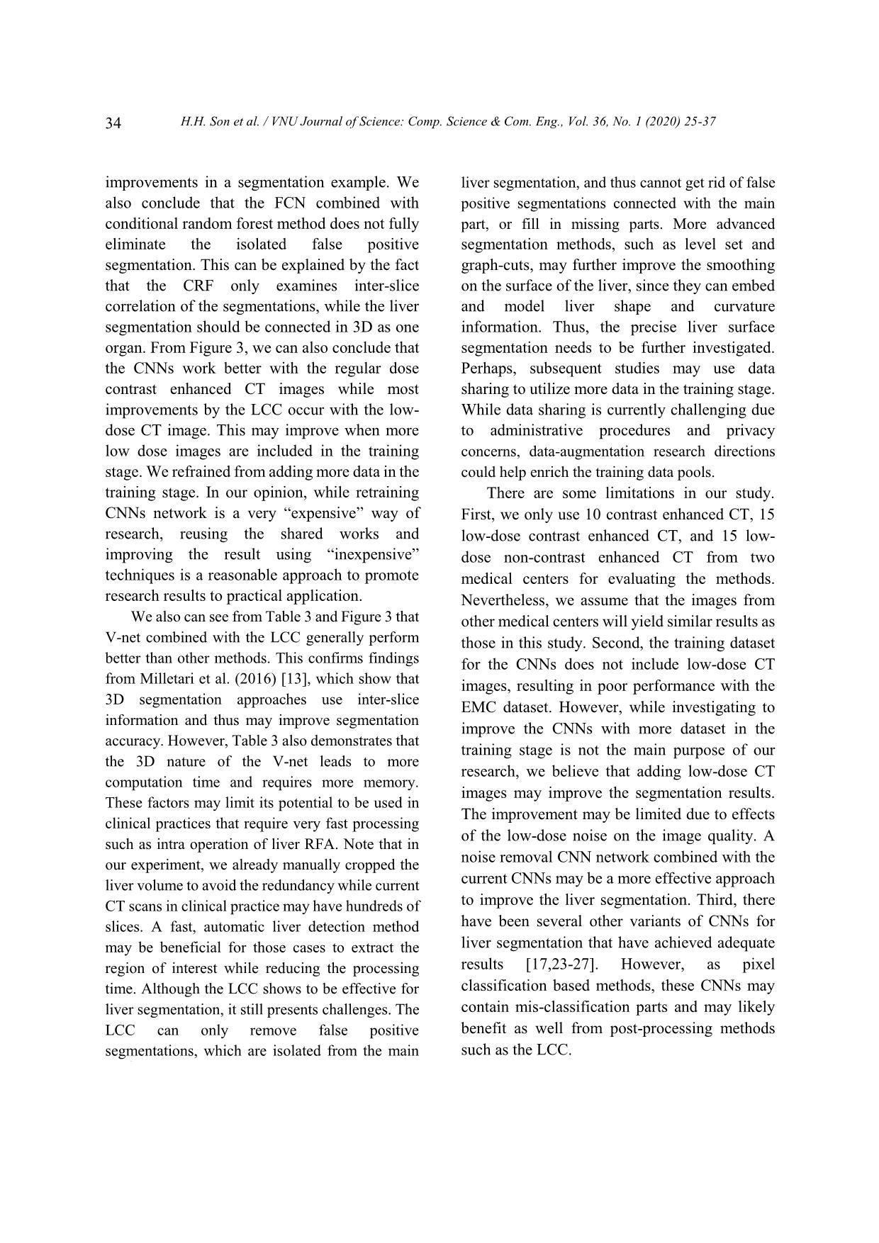
components method. The p-values of the t-tests
for the evaluations scores of the pairs
FM/FM_LC, DM/DM_LC, VM/VM_LV,
FEL/FEL_LC, DEL/DEL_LC, VEL/VEL_LC,
PEN/PEN_LC, DEN/DEN_LC and
VEN/VEN_LC are summarized in Table 4.
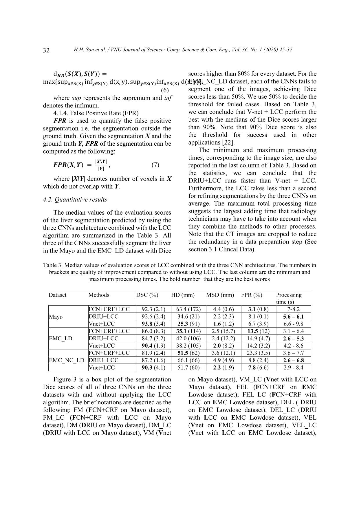
From Table 4, we can conclude that the LCC
algorithm statistically significantly improves the
segmentation results of all three CNNs
in general.
Table 4. P-values of the T-tests for the proposed method with the corresponding original CNNs:
The numbers are smaller than 0.05 indicating that the improvements are statistically significance.
Dataset Methods DSC HD MSD FPR
 FM/FM_LC 0.021 0.019 0.002 0.001
Mayo
DM/DM_LC 0.002 < 10
-3
< 10
-3
< 10
-3
 VM/VM_LC 0.040 0.001 0.014 0.019
 FEL/FEL_LC 0.010 < 10
-3
< 10
-3
< 10
-3
EMC_LD DEL/DEL_LC 0.016 < 10
-3
< 10
-3
0.118
 VEL/VEL_LC 0.027 < 10
-3
< 10
-3
< 10
-3
 FEN/FEN_LC 0.034 < 10-3 < 10-3 < 10-3
EMC_NC_LD DEN/DEN_LC 0.055 < 10
-3
< 10
-3
< 10
-3
 VEN/VEN_LC 0.019 < 10-3 < 10-3 < 10-3
p
The Figure 4 is an example of 3D liver
segmentations on a low-dose contrast enhanced
CT image. In the second column, the liver
segmentations by three CNNs include some false
positive segmentations (in blue), which are
eliminated by the LCC algorithm. Obviously, the
difference in segmentation from three networks
is not visible in the 2D view (right column). The
3D view in the first column visualizes the
difference between the liver segmentations and
the ground truth.
5. Discussion
In this study, we investigate the
improvement in liver segmentation using CNNs
approaches on CT images when they are
combined with a connected component
algorithm and the largest component in a post-
processing step. We either re-implement or reuse
the CNNs model trained with the LiTS dataset,
testing them with other three datasets from two
different medical centers with both standard and
low dose protocols with and without contrast
enhancement. Next, we apply the LCC algorithm
on the liver segmentations by the CNNs
approaches and quantitatively evaluate the
results using well-known criteria for liver
segmentation. Combination of the CNN
approaches with the LCC algorithm statistically
significantly improves the liver segmentation.
The 3D visualization in the Figure 4 shows the
H.H. Son et al. / VNU Journal of Science: Comp. Science & Com. Eng., Vol. 36, No. 1 (2020) 25-37
34
improvements in a segmentation example. We
also conclude that the FCN combined with
conditional random forest method does not fully
eliminate the isolated false positive
segmentation. This can be explained by the fact
that the CRF only examines inter-slice
correlation of the segmentations, while the liver
segmentation should be connected in 3D as one
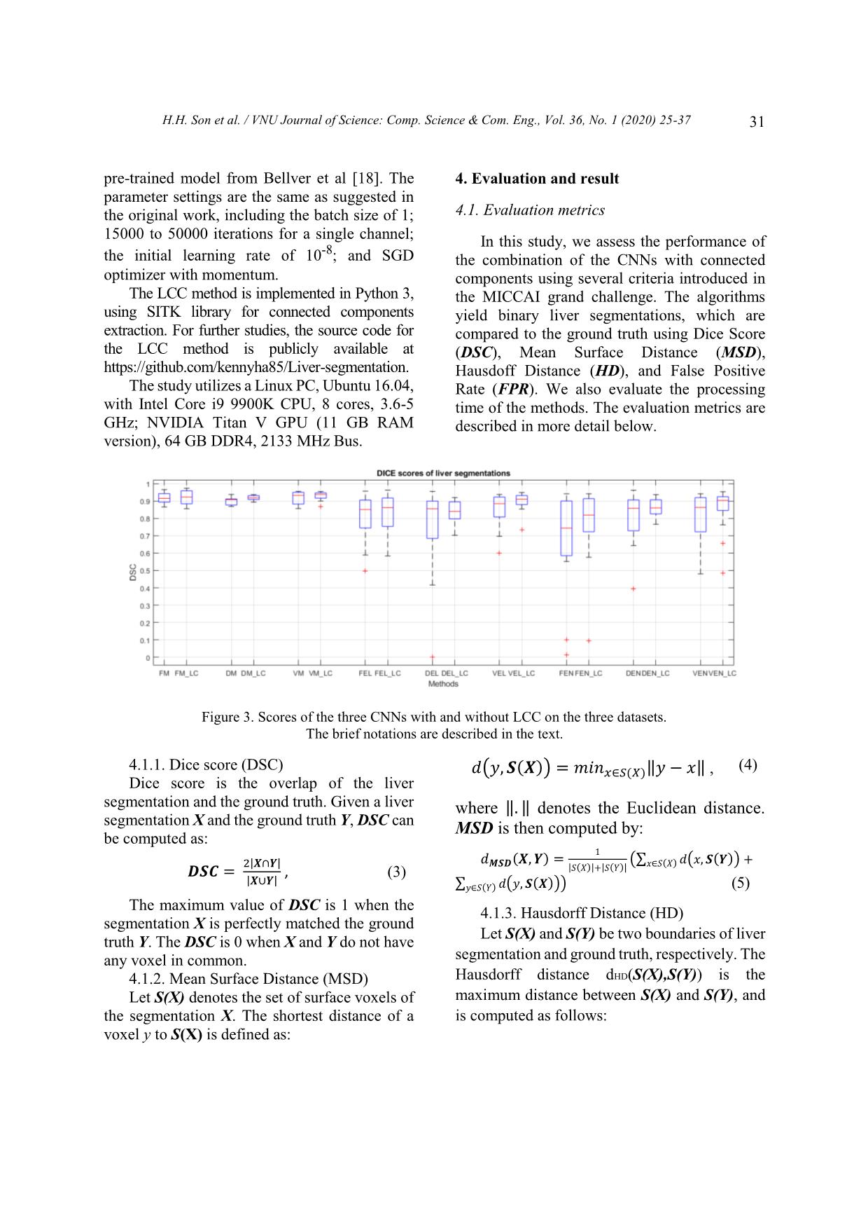
organ. From Figure 3, we can also conclude that
the CNNs work better with the regular dose
contrast enhanced CT images while most
improvements by the LCC occur with the low-
dose CT image. This may improve when more
low dose images are included in the training
stage. We refrained from adding more data in the
training stage. In our opinion, while retraining
CNNs network is a very “expensive” way of
research, reusing the shared works and
improving the result using “inexpensive”
techniques is a reasonable approach to promote
research results to practical application.
We also can see from Table 3 and Figure 3 that
V-net combined with the LCC generally perform
better than other methods. This confirms findings
from Milletari et al. (2016) [13], which show that
3D segmentation approaches use inter-slice
information and thus may improve segmentation
accuracy. However, Table 3 also demonstrates that
the 3D nature of the V-net leads to more
computation time and requires more memory.
These factors may limit its potential to be used in
clinical practices that require very fast processing
such as intra operation of liver RFA. Note that in
our experiment, we already manually cropped the
liver volume to avoid the redundancy while current
CT scans in clinical practice may have hundreds of
slices. A fast, automatic liver detection method
may be beneficial for those cases to extract the
region of interest while reducing the processing
time. Although the LCC shows to be effective for
liver segmentation, it still presents challenges. The
LCC can only remove false positive
segmentations, which are isolated from the main
liver segmentation, and thus cannot get rid of false
positive segmentations connected with the main
part, or fill in missing parts. More advanced
segmentation methods, such as level set and
graph-cuts, may further improve the smoothing
on the surface of the liver, since they can embed
and model liver shape and curvature
information. Thus, the precise liver surface
segmentation needs to be further investigated.
Perhaps, subsequent studies may use data
sharing to utilize more data in the training stage.
While data sharing is currently challenging due
to administrative procedures and privacy
concerns, data-augmentation research directions
could help enrich the training data pools.
There are some limitations in our study.
First, we only use 10 contrast enhanced CT, 15
low-dose contrast enhanced CT, and 15 low-
dose non-contrast enhanced CT from two
medical centers for evaluating the methods.
Nevertheless, we assume that the images from
other medical centers will yield similar results as
those in this study. Second, the training dataset
for the CNNs does not include low-dose CT
images, resulting in poor performance with the
EMC dataset. However, while investigating to
improve the CNNs with more dataset in the
training stage is not the main purpose of our
research, we believe that adding low-dose CT
images may improve the segmentation results.
The improvement may be limited due to effects
of the low-dose noise on the image quality. A
noise removal CNN network combined with the
current CNNs may be a more effective approach
to improve the liver segmentation. Third, there
have been several other variants of CNNs for
liver segmentation that have achieved adequate
results [17,23-27]. However, as pixel
classification based methods, these CNNs may
contain mis-classification parts and may likely
benefit as well from post-processing methods
such as the LCC.
F
H.H. Son et al. / VNU Journal of Science: Comp. Science & Com. Eng., Vol. 36, No. 1 (2020) 25-37
35
Figure 4. Example of 3D liver segmentations by the three CNNs on a low-dose contrast enhanced CT image.
The first raw is segmentations by FCN, the second one is by DRIU and the last one is by V-net. The first column
contains the liver segmentations using with LCC (green) and the ground truth (red), the second column illustrates
the raw liver segmentation from the CNNs (blue) overlapped by the segmentation after post processing, and the
last column is the final 2D liver segmentations on 2D CT slice of the liver.
6. Conclusion
In this paper, we present our work on
improving liver segmentation for CNN based
approaches using LCC algorithm. Experiments
are performed with three well-known CNN
architectures and with retrained or reused trained
models. We evaluate three datasets from two
different medical centers with regular contrast
enhanced CT image and both contrast and non-
contrast enhancement of low-dose image. The
quantitative evaluation results show that LCC
statistically significantly improves the liver
segmentation accuracy of the CNNs, while
maintaining the processing time of less than 10
seconds in total for all of the networks, including
the LCC processing time of less than a second.
In our study, we find that V-net combined with
the LCC achieves a Dice score of approximately
94%, which is comparable to other state of the
H.H. Son et al. / VNU Journal of Science: Comp. Science & Com. Eng., Vol. 36, No. 1 (2020) 25-37
36
art methods. We believe that with the current
development of CNN-based approach research, the
liver segmentation using CNNs has a high potential
to be applied in the clinical practice soon.
Acknowledgments
This work has been supported by VNU
University of Engineering and Technology
under project number CN 18.03. We would like
to thank Mayo Clinical for supporting us their
data. We also would like to thank NVIDIA for
their aid of a graphics hardware unit.
Reference
[1] K.A. McGlynn, J.L. Petrick, W.T. London, Global
epidemiology of hepatocellular carcinoma: An
emphasis on demographic and regional variability.
Clinics in liver disease 19(2) (2015)
223-238.
[2] M. Mohammadian, N. Mahdavifar, A.
Mohammadian-Hafshejani, H. Salehiniya, Liver
cancer in the world: epidemiology, incidence,
mortality and risk factors, World Cancer Res J.
5(2) (2018) e1082.
[3] T.T. Hong, N. Phuong Hoa, S.M. Walker, P.S. Hill,
C. Rao, Completeness and reliability of mortality
data in Viet Nam: Implications for the national
routine health management information system,
PloS one 13(1) 2018) e0190755.
https://doi.org/10.1371/journal.pone.0190755.
[4] T. Pham, L. Bui, G. Kim, D. Hoang, T. Tran,
M. Hoang, Cancers in Vietnam-Burden and
Control Efforts: A Narrative Scoping Review.
Cancer Control 26(1) (2019) 1073274819863802.
[5] M. Borner, M. Castiglione, J. Triller, H.U. Baer, M.
Soucek, L. Blumgart, K. Brunner, Arena:
Considerable side effects of chemoembolization
for colorectal carcinoma metastatic to the liver,
Annals of oncology 3(2) (1992) 113-115.
[6] K. Memon, R.J. Lewandowski, L. Kulik, A. Riaz,
M.F. Mulcahy, R. Salem, Radioembolization for
primary and metastatic liver cancer, In Seminars in
radiation oncology, WB Saunders. 21(4) (2011)
294-302.
[7] I. Gory, M. Fink, S. Bell, P. Gow, A. Nicoll, V.
Knight, W. Kemp, Radiofrequency ablation versus
resection for the treatment of early stage
hepatocellular carcinoma: A multicenter
Australian study, Scandinavian journal of
gastroenterology 50(5) (2015) 567-576.
[8] H.M. Luu, C. Klink, W. Niessen, A. Moelker, T.
Van Walsum, Non-rigid registration of liver CT
images for CT-guided ablation of liver tumors.
PloS one, 11(9) 92016) e0161600.
[9] G. Gunay, M.H. Luu, A. Moelker, T. Van Walsum,
S. Klein, Semiautomated registration of pre‐and
intraoperative CT for image‐guided percutaneous
liver tumor ablation interventions, Medical physics
44(7) (2017) 3718-3725.
[10] A. Gotra, L. Sivakumaran, G. Chartrand, N. Vu, F.
Vandenbroucke-Menu, C. Kauffmann, A. Tang,
Liver segmentation: indications, techniques and
future directions, Insights into imaging 8(4) (2017)
377-392. https://doi.org/10.1007/s13244-017-
0558-1.
[11] T. Heimann, B. Van Ginneken, M.A. Styner, Y.
Arzhaeva, V. Aurich, C. Bauer, F. Bello,
Comparison and evaluation of methods for liver
segmentation from CT datasets, IEEE transactions
on medical imaging 28(8) (2009) 1251-1265.
[12] O. Ronneberger, P. Fischer, T. Brox, U-net:
Convolutional networks for biomedical image
segmentation, In International Conference on
Medical image computing and computer-assisted
intervention, Springer, Cham, 2015, pp. 234-241.
[13] F. Milletari, N. Navab, S.A. Ahmadi, October,
V-net: Fully convolutional neural networks for
volumetric medical image segmentation, In 2016
Fourth International Conference on 3D Vision
(3DV) IEEE, 2016, pp. 565-571.
[14] P.F. Christ, F. Ettlinger, F. Grün, M.A. Elshaera, J.
Lipkova, S. Schlecht, M. Rempfler, Automatic
liver and tumor segmentation of CT and MRI
volumes using cascaded fully convolutional neural
networks, arXiv preprint arXiv:1702.05970, 2017.
[15] P.F. Christ, M.E.A. Elshaer, F. Ettlinger, S.
Tatavarty, M. Bickel, P. Bilic, H. Sommer,
Automatic liver and lesion segmentation in CT
using cascaded fully convolutional neural
networks and 3D conditional random fields. In
International Conference on Medical Image
Computing and Computer-Assisted Intervention,
Springer, Cham, 2016, pp. 415-423.
[16] H. Meine, G. Chlebus, M. Ghafoorian, I. Endo, A.
Schenk, Comparison of U-net-based
Convolutional Neural Networks for Liver
Segmentation in CT. arXiv preprint arXiv, 2018,
pp. 1810.04017.
[17] X. Li, H. Chen, X. Qi, Q. Dou, C.W. Fu, P.A.
Heng, H-DenseUNet: hybrid densely connected
H.H. Son et al. / VNU Journal of Science: Comp. Science & Com. Eng., Vol. 36, No. 1 (2020) 25-37
37
UNet for liver and tumor segmentation from CT
volumes, IEEE transactions on medical imaging,
37(12) (2018) 2663-2674.
[18] M. Bellver, K.K. Maninis, J. Pont-Tuset, X. Giró-
i-Nieto, J. Torres, L. Van Gool, Detection-aided
liver lesion segmentation using deep learning,
ArXiv preprint arXiv:1711.11069, 2017.
[19] H.S. Hoang, C.P. Pham, D. Franklin, T. Van
Walsum, M.H. Luu, An Evaluation of CNN-based
Liver Segmentation Methods using Multi-types of
CT Abdominal Images from Multiple Medical
Centers, In 2019 19th International Symposium on
Communications and Information Technologies
(ISCIT), IEEE, September, 2019, pp. 20-25.
[20] H. Samet, M. Tamminen, Efficient component
labeling of images of arbitrary dimension
represented by linear bintrees, IEEE Transactions
on Pattern Analysis and Machine Intelligence,
10(4) (1988) 579-586.
[21] P. Bilic, P.F. Christ, E. Vorontsov, G. Chlebus, H.
Chen, Q. Dou, S. Kadoury, The liver tumor
segmentation benchmark (lits), ArXiv preprint
arXiv, 2019, 1901.04056.
[22] H.M. Luu, A. Moelker, S. Klein, W. Niessen, T.
Van Walsum, Quantification of nonrigid liver
deformation in radiofrequency ablation
interventions using image registration, Physics in
Medicine & Biology 63(17) (2018) 175005.
[23] A.A. Novikov, D. Major, M. Wimmer, D. Lenis, K.
Bühler, Deep Sequential Segmentation of Organs
in Volumetric Medical Scans, IEEE transactions on
medical imaging, 2018.
[24] Y. Huo, J.G. Terry, J. Wang, S. Nair, A. Lasko, B.I.
Freedman, B.A. Landman, Fully Automatic Liver
Attenuation Estimation combing CNN
Segmentation and Morphological Operations,
Medical physics, 2019.
[25] N. Gruber, S. Antholzer, W. Jaschke, C. Kremser,
M. Haltmeier, A Joint Deep Learning Approach for
Automated Liver and Tumor Segmentation, ArXiv
preprint arXiv, 2019, pp. 1902.07971.
[26] S. Chen, K. Ma, Y. Zheng, Med3D: Transfer
Learning for 3D Medical Image Analysis, ArXiv
preprint arXiv, 2019, pp. 1904.00625.
[27] W. Tang, D. Zou, S. Yang, J. Shi, DSL: Automatic
Liver Segmentation with Faster
R-CNN and DeepLab, In International Conference
on Artificial Neural Networks, Springer, Cham,
2018, pp. 137-147.
O
-
File đính kèm:
 liver_segmentation_on_a_variety_of_computed_tomography_ct_im.pdf

